

Physical interaction between the herpes simplex virus type 1 exonuclease, UL12, and the DNA double-strand break-sensing MRN complex
- PMID: 20943970
- PMCID: PMC3004347
- DOI: 10.1128/JVI.01506-10
Physical interaction between the herpes simplex virus type 1 exonuclease, UL12, and the DNA double-strand break-sensing MRN complex
Abstract
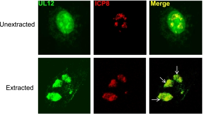
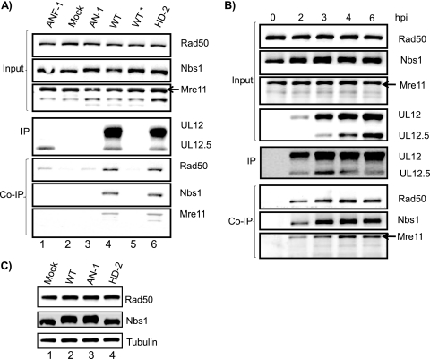
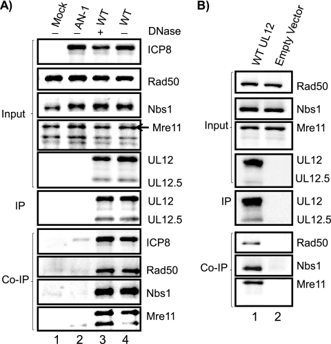
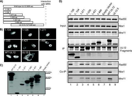
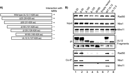
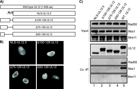
The herpes simplex virus type 1 (HSV-1) alkaline nuclease, encoded by the UL12 gene, plays an important role in HSV-1 replication, as a UL12 null mutant displays a severe growth defect. The HSV-1 alkaline exonuclease UL12 interacts with the viral single-stranded DNA binding protein ICP8 and promotes strand exchange in vitro in conjunction with ICP8. We proposed that UL12 and ICP8 form a two-subunit recombinase reminiscent of the phage lambda Red α/β recombination system and that the viral and cellular recombinases contribute to viral genome replication through a homologous recombination-dependent DNA replication mechanism. To test this hypothesis, we identified cellular interaction partners of UL12 by using coimmunoprecipitation. We report for the first time a specific interaction between UL12 and components of the cellular MRN complex, an important factor in the ATM-mediated homologous recombination repair (HRR) pathway. This interaction is detected early during infection and does not require viral DNA or other viral or cellular proteins. The region of UL12 responsible for the interaction has been mapped to the first 125 residues, and coimmunoprecipitation can be abolished by deletion of residues 100 to 126. These observations support the hypothesis that cellular and viral recombination factors work together to promote efficient HSV-1 growth.
Figures
Similar articles
-
HSV-1 DNA Replication-Coordinated Regulation by Viral and Cellular Factors.Viruses. 2021 Oct 7;13(10):2015. doi: 10.3390/v13102015. Viruses. 2021. PMID: 34696446 Free PMC article. Review.
-
The HSV-1 exonuclease, UL12, stimulates recombination by a single strand annealing mechanism.PLoS Pathog. 2012;8(8):e1002862. doi: 10.1371/journal.ppat.1002862. Epub 2012 Aug 9. PLoS Pathog. 2012. PMID: 22912580 Free PMC article.
-
The herpes simplex virus alkaline nuclease is required to maintain replication fork progression.J Virol. 2024 Dec 17;98(12):e0183624. doi: 10.1128/jvi.01836-24. Epub 2024 Nov 7. J Virol. 2024. PMID: 39508568 Free PMC article.
-
The Exonuclease Activity of Herpes Simplex Virus 1 UL12 Is Required for Production of Viral DNA That Can Be Packaged To Produce Infectious Virus.J Virol. 2017 Nov 14;91(23):e01380-17. doi: 10.1128/JVI.01380-17. Print 2017 Dec 1. J Virol. 2017. PMID: 28956767 Free PMC article.
-
The role of DNA recombination in herpes simplex virus DNA replication.IUBMB Life. 2003 Aug;55(8):451-8. doi: 10.1080/15216540310001612237. IUBMB Life. 2003. PMID: 14609200 Review.
Cited by
-
Activation of H2AX and ATM in varicella-zoster virus (VZV)-infected cells is associated with expression of specific VZV genes.Virology. 2014 Mar;452-453:52-8. doi: 10.1016/j.virol.2013.12.039. Epub 2014 Jan 29. Virology. 2014. PMID: 24606682 Free PMC article.
-
Herpes simplex viruses: mechanisms of DNA replication.Cold Spring Harb Perspect Biol. 2012 Sep 1;4(9):a013011. doi: 10.1101/cshperspect.a013011. Cold Spring Harb Perspect Biol. 2012. PMID: 22952399 Free PMC article. Review.
-
HSV-1 DNA Replication-Coordinated Regulation by Viral and Cellular Factors.Viruses. 2021 Oct 7;13(10):2015. doi: 10.3390/v13102015. Viruses. 2021. PMID: 34696446 Free PMC article. Review.
-
Replication and recombination of herpes simplex virus DNA.J Biol Chem. 2011 May 6;286(18):15619-24. doi: 10.1074/jbc.R111.233981. Epub 2011 Mar 1. J Biol Chem. 2011. PMID: 21362621 Free PMC article. Review.
-
DNA mismatch repair proteins are required for efficient herpes simplex virus 1 replication.J Virol. 2011 Dec;85(23):12241-53. doi: 10.1128/JVI.05487-11. Epub 2011 Sep 28. J Virol. 2011. PMID: 21957315 Free PMC article.
References
-
- Bataille, D., and A. Epstein. 1994. Herpes simplex virus replicative concatemers contain L components in inverted orientation. Virology 203:384-388. - PubMed
-
- Boehmer, P. E., and I. R. Lehman. 1997. Herpes simplex virus DNA replication. Annu. Rev. Biochem. 66:347-384. - PubMed
-
- Brown, S. M., J. H. Subak-Sharpe, J. Harland, and A. R. MacLean. 1992. Analysis of intrastrain recombination in herpes simplex virus type 1 strain 17 and herpes simplex virus type 2 strain HG52 using restriction endonuclease sites as unselected markers and temperature-sensitive lesions as selected markers. J. Gen. Virol. 73:293-301. - PubMed
Publication types
MeSH terms
Substances
Grants and funding
LinkOut - more resources
Full Text Sources
Other Literature Sources
Medical
Molecular Biology Databases
Research Materials
Miscellaneous
