. 2010 Dec;84(24):12934-44.
doi: 10.1128/JVI.01377-10. Epub 2010 Oct 13.

Cytoplasmic poly(A) binding proteins regulate telomerase activity and cell growth in human papillomavirus type 16 E6-expressing keratinocytes

Affiliations

Cytoplasmic poly(A) binding proteins regulate telomerase activity and cell growth in human papillomavirus type 16 E6-expressing keratinocytes

Rachel A Katzenellenbogen et al. J Virol. 2010 Dec.

Abstract

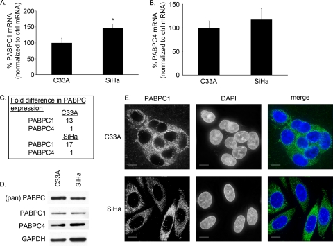
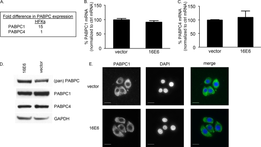
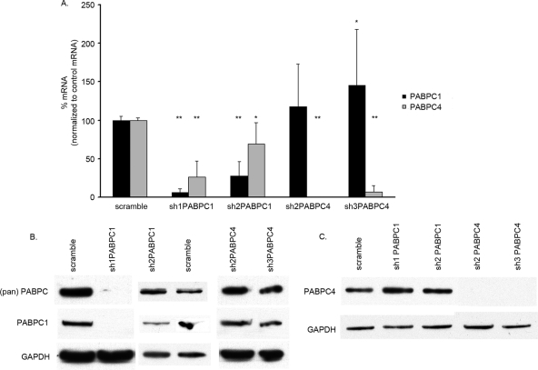
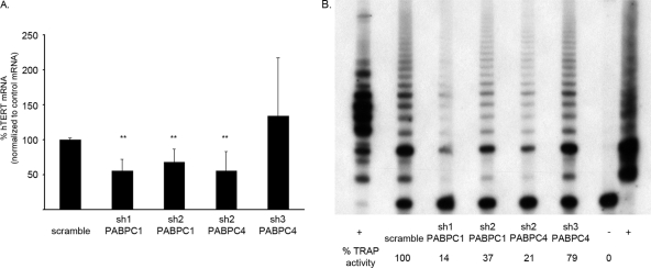
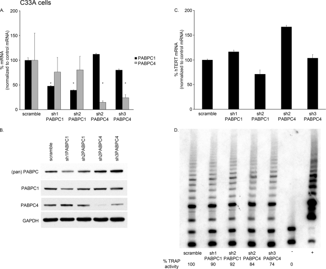
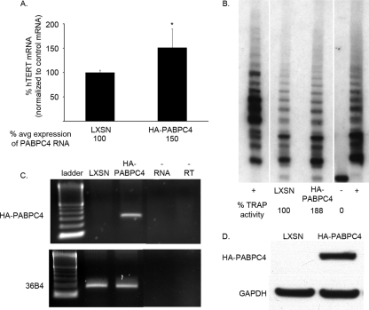
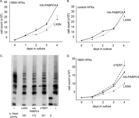
The high-risk human papillomavirus (HPV) E6 and E7 oncoproteins are critical to the immortalization of keratinocytes. HPV type 16 (HPV16) E6 interacts with endogenous proteins to activate hTERT, the catalytic subunit of telomerase, thus avoiding cellular senescence signals. NFX1-123, the longer splice variant of NFX1, interacts with HPV16 E6, as well as cytoplasmic poly(A) binding proteins 1 and 4 (PABPC1 and PABPC4). HPV16 E6 affects hTERT expression posttranscriptionally through NFX1-123, as NFX1-123 interacts with hTERT mRNA and stabilizes it, leading to greater telomerase activity. The PAM2 motif of NFX1-123, with which it binds PABPCs, is required for the posttranscriptional regulation of hTERT by HPV16 E6 and NFX1-123. There is increasing evidence that RNA and DNA viruses utilize RNA-processing proteins, and specifically PABPCs, in the normal virus life cycle, and there is also evidence that RNA-processing proteins are perturbed in cancers. Here, we show that PABPCs are critical in hTERT regulation by HPV16 E6. Although the amount and cellular localization of PABPCs were largely unchanged in cervical cancer cell lines with or without HPV16 and in human foreskin keratinocytes (HFKs) with or without HPV16 E6, knockdown of PABPCs decreased hTERT mRNA and telomerase activity and overexpression of PABPC4 increased these in HPV16 E6-expressing HFKs. In contrast, knockdown of PABPCs in C33A cells had no effect on hTERT mRNA or telomerase activity. Additionally, overexpression of PABPC4 and hTERT led to greater growth of cultured HPV16 E6-expressing HFKs. This is the first evidence that PABPCs have a targeted role in hTERT regulation leading to a growth advantage in cells expressing HPV16 E6.


Figures

FIG. 1.
FIG. 1.
PABPC1 and PABPC4 were expressed in HPV16-negative and -positive cell lines C33A and SiHa. (A to C) mRNA levels were detected by qPCR, normalized to the level of mRNA for 36B4, an endogenous ribosomal protein (control [ctrl]), and then expressed as a ratio. (A) PABPC1 mRNA levels were detected by qPCR, and in SiHa cells PABPC1 was expressed at 150% the amount found in C33A cells (*, P = 0.01; error bars indicate 95% CI). (B) PABPC4 mRNA levels were detected by qPCR and found to be similar in C33A and SiHa cells (P = 0.29; error bars indicate 95% CI). (C) PABPC1 and PABPC4 mRNAs were expressed in C33A and SiHa cells at ratios of 13:1 and 17:1, respectively. (D) Western blot analysis of whole-cell extracts showed equal levels of expression of all PABPCs and specifically PABPC1 and PABPC4 in C33A and SiHa cells. (pan) PABPC, antibody to region common to all PABPCs; PABPC1, antibody to unique region of PABPC1; PABPC4, antibody to unique region of PABPC4; GAPDH, loading control. (E) C33A and SiHa cells expressed PABPC1 primarily in the cytoplasm, although there was a minimal amount of PABPC1 found in the nuclei of SiHa cells, in contrast to C33A cells. (Green, PABPC1; blue, DAPI; white bars, 10 μm.)
FIG. 2.
FIG. 2.
PABPC1 and PABPC4 were expressed in HFKs with and without HPV16 E6. (A to C) mRNA levels were detected by qPCR, normalized to the level of mRNA for 36B4, an endogenous ribosomal protein (control [ctrl]), and then expressed as a ratio. (A) PABPC1 and PABPC4 were expressed in HFKs. Their relative mRNA expression levels were approximately 15:1. (B) PABPC1 mRNA levels were detected by qPCR and were similar in HFKs with and without HPV16 E6 (P = 0.14; error bars indicate 95% CI). (C) PABPC4 mRNA levels were detected by qPCR and were similar in HFKs with and without HPV16 E6 (P = 0.35; error bars indicate 95% CI). (D) Western blot analysis of whole-cell extracts showed equal levels of expression of all PABPCs and specifically PABPC1 and PABPC4 in HFKs with and without HPV16 E6. (pan) PABPC, antibody to region common to all PABPCs; PABPC1, antibody to unique region of PABPC1; PABPC4, antibody to unique region of PABPC4; GAPDH, loading control. (E) HFKs with and without HPV16 E6 expressed PABPC1 in the cytoplasm. (Green, PABPC1; blue, DAPI; white bars, 10 μm.)
FIG. 3.
FIG. 3.
PABPC1 and PABPC4 knockdown by shRNA in HPV16 E6-expressing HFKs. (A) PABPC1 and PABPC4 mRNA levels were detected by qPCR and graphed as percentages after normalization to levels in the presence of the scramble control. All P values were calculated by comparing results for an shRNA to results for the scramble control. sh1PABPC1 knocked down PABPC1 mRNA to 7% and PABPC4 to 27% (**, P < 0.0001 for both). sh2PABPC1 knocked down PABPC1 mRNA to 27% and PABPC4 to 69% (**, P < 0.0001; *, P = 0.0029). sh2PABPC4 knocked down PABPC4 to 1% (**, P < 0.0001). sh3PABPC4 knocked down PABPC4 mRNA to 8% and increased PABPC1 mRNA levels to 146% (**, P < 0.0001; *, P = 0.0421). These data are a summary of results from triplicate qPCRs conducted with three HPV16 E6-expressing HFK cell lines, with values normalized to 36B4 control mRNA levels. (Error bars, 95% CI.) (B) Western blot analysis of whole-cell extracts showed knockdown of PABPC1 by sh1PABPC1 and reduction of PABPC1 by sh2PABPC1 compared to levels in the presence of the scramble vector and a loading control. (pan) PABPC, antibody to region common to all PABPCs; PABPC1, antibody to unique region of PABPC1; GAPDH, loading control. (C) Western blot analysis of whole-cells extracts showed knockdown of PABPC4 by sh2PABPC4 and sh3PABPC4 compared to levels in the presence of the scramble shRNA and the loading control. PABPC4, antibody to unique region of PABPC4; GAPDH, loading control.
FIG. 4.
FIG. 4.
Knockdown of PABPC1 or PABPC4 decreased hTERT expression in HPV16 E6-expressing HFKs. (A) shRNA expressed in HPV16 E6-expressing HFKs knocked down PABPC1, PABPC4, or both. When PABPC1, PABPC4, or both were reduced by shRNA, hTERT mRNA levels were also reduced relative to those in the presence of the scramble control. When PABPC4 was reduced but PABPC1 was increased, there was no difference in hTERT mRNA levels. These data are a summary of results from triplicate qPCRs conducted with three HPV16 E6-expressing HFK cell lines, with values normalized to 36B4 control mRNA levels. (**, P ≤ 0.002; error bars indicate 95% CI.) (B) Telomerase activity in HPV16 E6-expressing HFKs was reduced when PABPC1, PABPC4, or both were knocked down by shRNA. These data are representative of results for three HPV16 E6-expressing HFK cell lines. (+, TSR8 and HeLa positive controls; −, negative control.)
FIG. 5.
FIG. 5.
Knockdown of PABPC1 or PABPC4 in C33A cells did not affect hTERT expression. (A) PABPC1 and PABPC4 mRNA levels were detected by qPCR and graphed as percentages after normalization to levels in the presence of the scramble control. sh1PABPC1 knocked down PABPC1 mRNA to 47%, and sh2PABPC1 knocked down PABPC1 mRNA to 39%. sh2PABPC4 knocked down PABPC4 to 15%, and sh3PABPC4 knocked down PABPC4 mRNA to 24%. sh2PABPC4 and sh3PABPC4 had minor effects on PABPC1 levels, and sh1PABPC1 and sh2PABPC1 had no effect on PABPC4 levels. These data are from triplicate qPCRs and were normalized to 36B4 control mRNA levels. All P values were calculated by comparing results for an shRNA to the results for the scramble control. (*, P ≤ 0.05; error bars, 95% CI.) (B) Western blot analysis of whole-cell extracts showed some decrease in PABPC1 by sh1PABPC1 and sh2PABPC1 and larger decreases in PABPC4 by sh2PABPC4 and sh3PABPC4 compared to levels in the presence of the scramble vector and a loading control. (pan) PABPC, antibody to region common to all PABPCs. PABPC1, antibody to unique region of PABPC1; PABPC4, antibody to unique region of PABPC4; GAPDH, loading control. (C) When PABPC1 or PABPC4 was knocked down by shRNA, hTERT mRNA levels were not consistently reduced relative to those in the presence of the scramble control. Error bars, 95% CI. (D) Telomerase activity in C33A cells was not consistently reduced when PABPC1 or PABPC4 was knocked down by shRNA. These data are representative of results from three separate experiments. (+, HeLa positive control; −, negative control.)
FIG. 6.
FIG. 6.
Overexpression of PABPC4 increased hTERT expression in HPV16 E6-expressing HFKs. (A) HA-tagged PABPC4 expressed in HPV16 E6-expressing HFKs increased total PABPC4 mRNA levels to 150% of endogenous levels by qPCR. Overexpression of PABPC4 increased hTERT to 150% that found in LXSN (vector control) HPV16 E6-expressing cells. These data are a summary of results from triplicate qPCRs conducted with three HPV16 E6-expressing HFK cell lines, with values normalized to 36B4 control mRNA levels. (*, P < 0.05; error bars indicate 95% CI.) (B) Telomerase activity in HPV16 E6-expressing HFKs overexpressing HA-tagged PABPC4 was increased relative to that in LXSN- and HPV16 E6-expressing cells. These data are representative of results for three HPV16 E6-expressing HFK cell lines. (+, TSR8 and HeLa positive controls; −, negative control.) (C) HA-tagged PABPC4 was detected by RT-PCR. (36B4, positive RNA control; −RNA, no RNA template; −RT, no reverse transcriptase.) (D) HA-tagged PABPC4 was detected by Western blot analysis of whole-cell extracts. (GAPDH, loading control.)
FIG. 7.
FIG. 7.
Overexpression of PABPC4 and hTERT increased daily growth of HPV16 E6-expressing HFKs in culture. (A) HPV16 E6-expressing HFKs overexpressing HA-tagged PABPC4 and LXSN (vector control)/HPV16 E6-expressing cells were plated in equal numbers and counted daily for 4 days. HPV16 E6-expressing HFKs overexpressing HA-PABPC4 grew to greater numbers over 4 days. (Error bars, 95% CI.) (B) pBABEpuro HFKs (control HFKs) overexpressing HA-PABPC4 and LXSN control HFKs were plated in equal numbers and counted daily for 4 days. Control HFKs overexpressing HA-PABPC4 did not grow to greater numbers over 4 days. (Error bars, 95% CI.) (A and B) These data are representative of results for two different HPV16 E6- and pBABEpuro-expressing HFK cell lines counted in triplicate in two different experiments. (C) Telomerase activity in HPV16 E6-expressing HFKs overexpressing HA-tagged PABPC4 or hTERT was increased relative to that in LXSN/HPV16 E6-expressing cells. (+, TSR8 and HeLa positive controls; −, negative control.) (D) HPV16 E6-expressing HFKs overexpressing HA-PABPC4 or hTERT and LXSN (vector control)/HPV16 E6-expressing cells were plated in equal numbers and counted daily for 4 days. HPV16 E6-expressing HFKs overexpressing HA-PABPC4 grew as well as those overexpressing hTERT, and both grew to greater numbers than LXSN/HPV16 E6-expressing cells. (Error bars, 95% CI.) These data are representative of results for two HPV16 E6-expressing cell lines counted in triplicate.

Similar articles

Cited by

References

    1. Albrecht, M., and T. Lengauer. 2004. Survey on the PABC recognition motif PAM2. Biochem. Biophys. Res. Commun. 316:129-138. - PubMed
    1. Amrani, N., S. Ghosh, D. A. Mangus, and A. Jacobson. 2008. Translation factors promote the formation of two states of the closed-loop mRNP. Nature 453:1276-1280. - PMC - PubMed
    1. Auersperg, N. 1964. Long-term cultivation of hypodiploid human tumor cells. J. Natl. Cancer Inst. 32:135-163. - PubMed
    1. Baker, C. C., W. C. Phelps, V. Lindgren, M. J. Braun, M. A. Gonda, and P. M. Howley. 1987. Structural and transcriptional analysis of human papillomavirus type 16 sequences in cervical carcinoma cell lines. J. Virol. 61:962-971. - PMC - PubMed
    1. Bartz, S. R., and M. A. Vodicka. 1997. Production of high-titer human immunodeficiency virus type 1 pseudotyped with vesicular stomatitis virus glycoprotein. Methods 12:337-342. - PubMed

Publication types

MeSH terms

LinkOut - more resources