. 2010 Dec;84(24):12492-503.
doi: 10.1128/JVI.01495-10. Epub 2010 Oct 13.

Nodavirus-induced membrane rearrangement in replication complex assembly requires replicase protein a, RNA templates, and polymerase activity

Affiliations

Nodavirus-induced membrane rearrangement in replication complex assembly requires replicase protein a, RNA templates, and polymerase activity

Benjamin G Kopek et al. J Virol. 2010 Dec.

Abstract

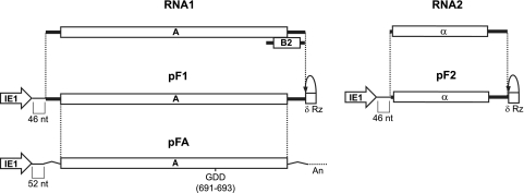
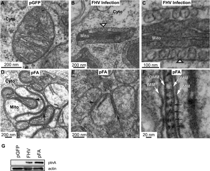
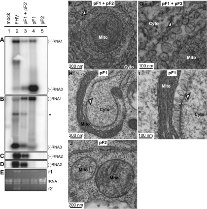
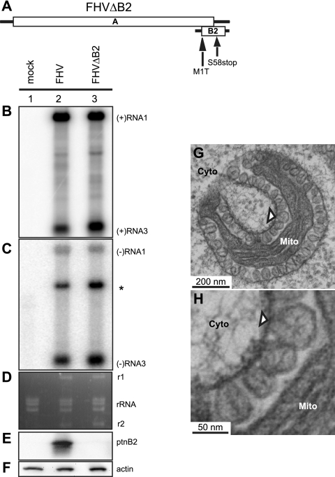
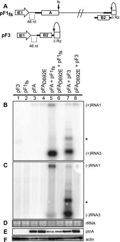
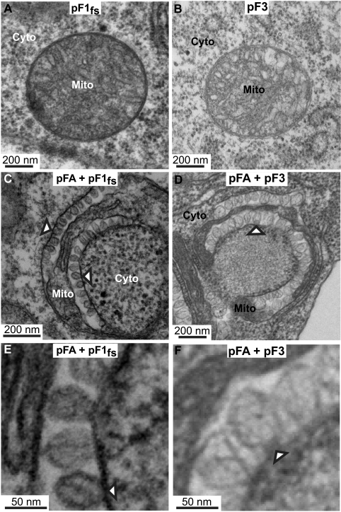
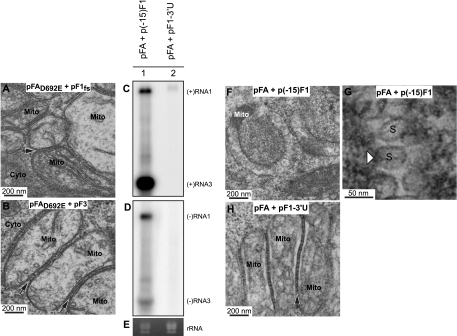
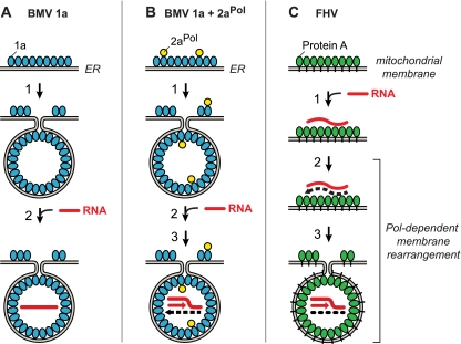
Positive-strand RNA [(+)RNA] viruses invariably replicate their RNA genomes on modified intracellular membranes. In infected Drosophila cells, Flock House nodavirus (FHV) RNA replication complexes form on outer mitochondrial membranes inside ∼50-nm, virus-induced spherular invaginations similar to RNA replication-linked spherules induced by many (+)RNA viruses at various membranes. To better understand replication complex assembly, we studied the mechanisms of FHV spherule formation. FHV has two genomic RNAs; RNA1 encodes multifunctional RNA replication protein A and RNA interference suppressor protein B2, while RNA2 encodes the capsid proteins. Expressing genomic RNA1 without RNA2 induced mitochondrial spherules indistinguishable from those in FHV infection. RNA1 mutation showed that protein B2 was dispensable and that protein A was the only FHV protein required for spherule formation. However, expressing protein A alone only "zippered" together the surfaces of adjacent mitochondria, without inducing spherules. Thus, protein A is necessary but not sufficient for spherule formation. Coexpressing protein A plus a replication-competent FHV RNA template induced RNA replication in trans and membrane spherules. Moreover, spherules were not formed when replicatable FHV RNA templates were expressed with protein A bearing a single, polymerase-inactivating amino acid change or when wild-type protein A was expressed with a nonreplicatable FHV RNA template. Thus, unlike many (+)RNA viruses, the membrane-bounded compartments in which FHV RNA replication occurs are not induced solely by viral protein(s) but require viral RNA synthesis. In addition to replication complex assembly, the results have implications for nodavirus interaction with cell RNA silencing pathways and other aspects of virus control.


Figures

FIG. 1.
FIG. 1.
Schematic of FHV genome components and the corresponding expression cassettes used in Drosophila expression plasmids. Each FHV component is expressed by a baculovirus IE1 promoter in a plasmid that also contains the baculovirus transactivating hr5 enhancer. Plasmid pF1 expresses RNA1, and pF2 expresses RNA2 with a δRz-mediated authentic 3′ end (δRz). pF1 and pF2 contain 46 nonviral nt between the IE1 transcription start site and the first viral nucleotide. pFA encodes protein A mRNA with nonviral 5′ and 3′ untranslated sequences and a 3′ polyadenylation signal (An). pFA contains 52 nonviral nt between the IE1 transcription start site and the initiating methionine codon.
FIG. 2.
FIG. 2.
Expression of protein A causes mitochondrial zippering. (A) Electron micrograph of a cell expressing GFP showing no apparent alterations in mitochondrial morphology. (B) Electron micrograph of a cell infected with WT FHV at 14 hpi showing formation of mitochondrial spherules (arrowhead) that represent the FHV RNA replication complex. (C) Higher-magnification view of spherules in panel B. (D and E) Electron micrograph of cells expressing pFA, showing mitochondrial aggregation and zippering (arrows) but no spherules. (F) Higher-magnification view of zippered mitochondria, showing structure between mitochondria that may be responsible for zippering effect (arrows). Mitochondria (Mito), cytoplasm (Cyto), inner mitochondrial membrane (IMM), and outer mitochondrial membrane (OMM) are indicated. (G) Total protein was analyzed by Western blotting with antibodies against protein A (ptnA; top) or actin (bottom).
FIG. 3.
FIG. 3.
Expression of replication-competent FHV RNA1 is sufficient to induce mitochondrial spherules. (A to E) Total RNA and total protein were isolated from Drosophila cells transfected with pGFP (mock) or pF1 and pF2 together or alone or infected with FHV. Total RNA was analyzed by Northern blotting with 32P-labeled cRNA probes for positive-strand RNA1 and RNA3 (A), negative-strand RNA1 and RNA3 (B), positive-strand RNA2 (C), and negative-strand RNA2 (D). (E) Ethidium bromide (EtBr)-stained rRNA is shown as a loading control. FHV RNA1 (r1) and RNA2 (r2) replicate to rRNA levels and can be observed by EtBr staining. The asterisk in panel B represents cross-reactivity of the negative-strand RNA1 probe with RNA2. The band that occurs between RNA1 and RNA3 in lane 2 of panel B may represent a defective interfering RNA (63) or an RNA2-RNA3 heterodimer (4). A similar band is also visible in Fig. 4B. (F) Electron micrograph of a cell expressing pF1 and pF2, showing the formation of mitochondrial spherules (arrowhead) similar to those observed for FHV infection (Fig. 2B and C). (G) Higher-magnification view of spherules in panel F. (H) Electron micrograph of a cell expressing pF1 alone, showing spherule formation (arrowhead). (I) Higher-magnification view of spherules in panel H. (J) Electron micrograph of a cell expressing pF2. Mitochondria (Mito) and cytoplasm (Cyto) are indicated. It should be noted that, as previously established by EM tomography, mitochondria bearing FHV spherules are frequently cup shaped, so that two-dimensional images of these mitochondria can appear quite different, depending on whether the plane of sectioning is perpendicular (Fig. 3F) or parallel (Fig. 3H) to the axis around which the mitochondrion is cupped. See Fig. 1 in reference for further explanation.
FIG. 4.
FIG. 4.
FHV protein B2 is not required for spherule formation. (A) Schematic of RNA1 showing mutations in the B2 ORF. Total RNA and total protein were isolated from Argonaute2 (ago2)-silenced Drosophila cells left uninfected (mock) or infected with WT FHV or FHV with mutations in the B2 ORF to block B2 expression (FHVΔB2). Total RNA was analyzed by Northern blotting with 32P-labeled cRNA probes for positive-strand RNA1 and RNA3 (B) or negative-strand RNA1 and RNA3 (C). The asterisk represents cross-reactivity of the negative-strand RNA1 probe with RNA2. (D) EtBr-stained rRNA is shown as a loading control and also shows that RNA1 (r1) and RNA2 (r2) are replicated to rRNA levels. Total protein was analyzed by Western blotting with antibodies against protein B2 (E) or actin (F) showing that cells infected with FHVΔB2 do not express detectable levels of protein B2. (G) Electron micrograph of ago2-silenced cells infected with FHVΔB2, showing spherule formation (arrowhead) in the absence of protein B2 expression. (H) Higher-magnification view of spherules in panel G. Mitochondria (Mito) and cytoplasm (Cyto) are indicated.
FIG. 5.
FIG. 5.
trans replication of FHV RNA templates. (A) Schematic of plasmid-directed RNA1fs and RNA3 expression in Drosophila cells. pF1fs is the same as pF1, except that a 4-nt sequence causes a frameshift (fs) that prevents full-length protein A production. pF3 expresses RNA3 from the IE1 promoter with a δRz-mediated authentic 3′ end (δRz). The 5′ ends of pF1fs and pF3 have 46 nonviral nt between the transcription start site and the first viral nucleotide, as was shown for pF1 and pF2. Drosophila cells were transfected with pF3, pF1fs, pFA, pFAD692E, pFA plus pF1fs, pFAD692E plus pF1fs, pFA plus pF3, or pFAD692E plus pF3. (B to D) Total RNA was analyzed by Northern blotting with 32P-labeled cRNA probes for positive-strand RNA1 and RNA3 (B) or negative-strand RNA1 and RNA3 (C). Note that in panel B generally low but somewhat varying levels of the expected pFA- or pFAD692E-generated, nonreplicatable protein A mRNA transcripts are visible in lanes 3 to 8 at a position slightly above the replicated positive-strand RNA1 band in lane 5. As noted in an earlier study (59), the similarly sized, weak band near RNA1 in panel C, lane 7, appears to represent detection of an incompletely denatured hybrid between negative-strand RNA3 and the positive-strand protein A mRNA. This band does not represent negative-strand RNA1 since, despite its high position in the gel, the band is only detected with a strand-specific probe against the common sequences of negative-strand RNA3 and RNA1, but not with a strand-specific probe against the unique 3′-proximal portion of negative-strand RNA1, outside of the sequences shared with RNA3 (59). (D) EtBr-stained rRNA is shown as a loading control. Total protein was analyzed by Western blotting with antibodies against protein A (E) or actin (F). The asterisk represents a slower-migrating form of RNA3 that has been previously observed.
FIG. 6.
FIG. 6.
Coexpressing FHV RNA templates with WT protein A induces mitochondrial spherules. (A) Electron micrograph of a cell expressing pF1fs alone, showing no mitochondrial spherules. (B) Electron micrograph of a cell expressing pF3 alone, showing no mitochondrial spherules. (C) Electron micrograph of a cell expressing pFA plus pF1fs in which trans replication of RNA1fs is occurring (Fig. 5, lane 5), showing mitochondrial spherule formation (arrowheads). (D) Electron micrograph of a cell expressing pFA plus pF3 in which trans replication of RNA3 is occurring (Fig. 5, lane 7), showing mitochondrial spherule formation (arrowheads). (E and F) Higher-magnification views of spherules from panels C and D, respectively. Mitochondria (Mito) and cytoplasm (Cyto) are indicated.
FIG. 7.
FIG. 7.
Spherule formation requires protein A polymerase activity and a replicatable RNA template. (A) Electron micrograph of a cell expressing pFAD692E plus pF1fs in which no replication is occurring (Fig. 5, lane 6), showing no mitochondrial spherule formation but mitochondrial zippering (arrow), as occurs when expressing pFA alone (Fig. 2D to F). (B) Electron micrograph of a cell expressing pFAD692E plus pF3 in which no replication is occurring (Fig. 5, lane 8), showing no mitochondrial spherule formation but mitochondrial zippering (arrows), as occurs in panel A and when expressing pFA alone (Fig. 2D to F). Drosophila cells were transfected with pFA plus an RNA1fs template with a truncation of the first 15 nt [p(−15)F1] or a deletion of the 3′ UTR [pF1-3′U]. Total RNA was analyzed by Northern blotting with 32P-labeled cRNA probes for positive-strand RNA1 and RNA3 (C) or negative-strand RNA1 and RNA3 (D). (E) EtBr-stained rRNA. (F) Electron micrograph of cells transfected with pFA plus p(−15)F1. (G) Higher-magnification view of spherules from panel F. (H) Electron micrograph of cells transfected with pFA plus pF1-3′U, showing mitochondrial zippering but no spherules. Mitochondria (Mito), cytoplasm (Cyto), and spherules (S) are indicated.
FIG. 8.
FIG. 8.
Polymerase independence and dependence of BMV and FHV spherule membrane rearrangements and replication complex assembly. (A) BMV spherule generation by multifunctional BMV RNA replication factor 1a (blue). In the absence of BMV RNA-dependent RNA polymerase 2aPol, 1a localizes to ER membranes, self-interacts, and induces the formation of ∼70-nm invaginations or spherules (39, 40, 51). In a separable subsequent reaction dependent on the activity of the C-proximal 1a NTPase/helicase domain, 1a transfers genomic RNA (red line) to the spherule interior (61). (B) In the presence of 1a and 2aPol (yellow), 1a also recruits 2aPol to ER membranes and directs spherule formation and genomic RNA recruitment as in panel A. 2aPol then synthesizes negative-strand RNA (dashed black line) that is retained in the spherule and repeatedly used as a template to synthesize new positive strands. (C) Protein A (green), the sole FHV-encoded RNA replication factor, localizes to mitochondrial outer membranes (34) and self-interacts (16) but does not induce spherule formation unless replication-competent FHV RNA templates are present and protein A's RNA polymerase domain is active (Fig. 2 to 7). Active-site mutations abolishing protein A polymerase activity or deletion of 3′ RNA replication signals still allow protein A to recognize specific 5′-proximal elements in viral RNA templates and recruit them to mitochondrial membranes (59, 60) but block RNA synthesis and invagination of FHV spherules (Fig. 7). See Discussion for additional details, including the role of FHV RNA interference suppressor B2.

Similar articles

Cited by

References

    1. Ahlquist, P. 2006. Parallels among positive-strand RNA viruses, reverse-transcribing viruses and double-stranded RNA viruses. Nat. Rev. Microbiol. 4:371-382. - PMC - PubMed
    1. Ahola, T., and P. Ahlquist. 1999. Putative RNA capping activities encoded by brome mosaic virus: methylation and covalent binding of guanylate by replicase protein 1a. J. Virol. 73:10061-10069. - PMC - PubMed
    1. Ahola, T., J. A. den Boon, and P. Ahlquist. 2000. Helicase and capping enzyme active site mutations in brome mosaic virus protein 1a cause defects in template recruitment, negative-strand RNA synthesis, and viral RNA capping. J. Virol. 74:8803-8811. - PMC - PubMed
    1. Albariño, C. G., B. D. Price, L. D. Eckerle, and L. A. Ball. 2001. Characterization and template properties of RNA dimers generated during Flock House virus RNA replication. Virology 289:269-282. - PubMed
    1. Aliyari, R., Q. Wu, H. W. Li, X. H. Wang, F. Li, L. D. Green, C. S. Han, W. X. Li, and S. W. Ding. 2008. Mechanism of induction and suppression of antiviral immunity directed by virus-derived small RNAs in Drosophila. Cell Host Microbe 4:387-397. - PMC - PubMed

Publication types

MeSH terms

LinkOut - more resources