. 2010 Dec;84(24):12646-57.
doi: 10.1128/JVI.01328-10. Epub 2010 Oct 13.

Interferon-induced cell membrane proteins, IFITM3 and tetherin, inhibit vesicular stomatitis virus infection via distinct mechanisms

Affiliations

Interferon-induced cell membrane proteins, IFITM3 and tetherin, inhibit vesicular stomatitis virus infection via distinct mechanisms

Jessica M Weidner et al. J Virol. 2010 Dec.

Abstract

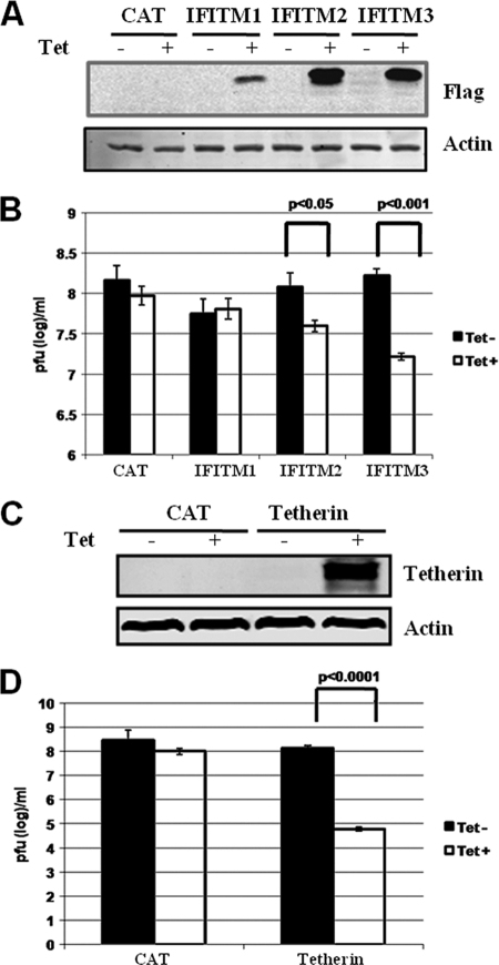
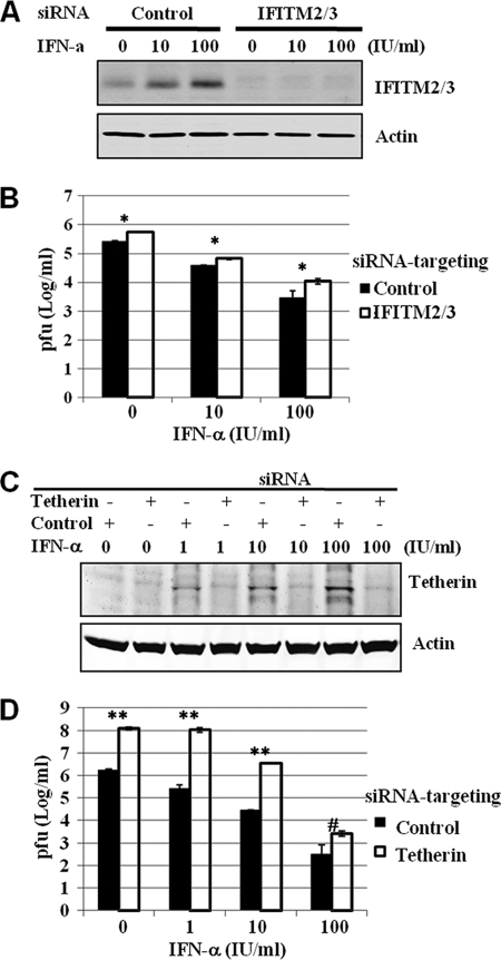
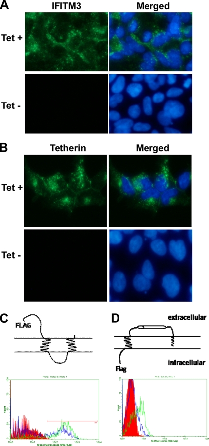
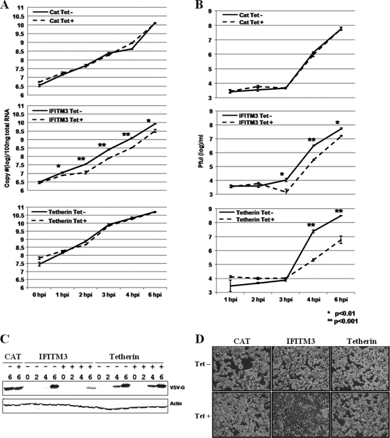
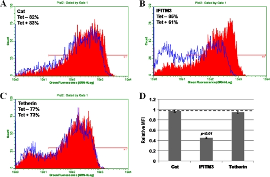
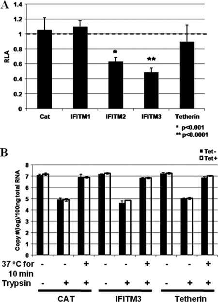
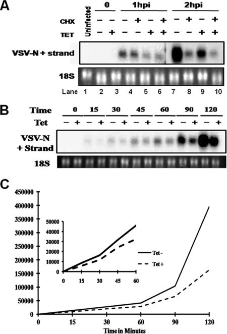
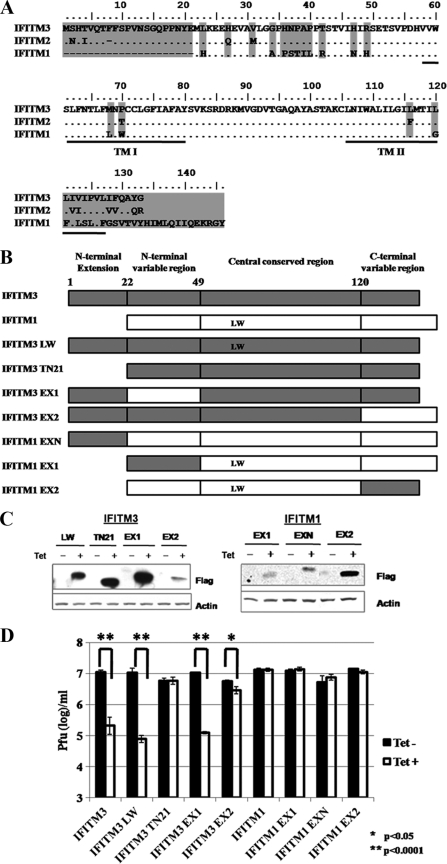
Tetherin and IFITM3 are recently identified interferon-induced cellular proteins that restrict infections by retroviruses and filoviruses and of influenza virus and flaviviruses, respectively. In our efforts to further explore their antiviral activities against other viruses and determine their antiviral mechanisms, we found that the two antiviral proteins potently inhibit the infection of vesicular stomatitis virus (VSV), a prototype member of the Rhabdoviridae family. Taking advantage of this well-studied virus infection system, we show that although both tetherin and IFITM3 are plasma membrane proteins, tetherin inhibits virion particle release from infected cells, while IFITM3 disrupts an early event after endocytosis of virion particles but before primary transcription of incoming viral genomes. Furthermore, we demonstrate that both the N-terminal 21 amino acid residues and C-terminal transmembrane region of IFITM3 are required for its antiviral activity. Collectively, our work sheds light on the mechanisms by which tetherin and IFITM3 restrict infection with rhabdoviruses and possibly other pathogenic viruses.


Figures

FIG. 1.
FIG. 1.
Antiviral activities of IFITMs and tetherin against VSV in FLP-IN T Rex cells, expressing CAT or the desired ISGs, left untreated or treated with 1 μg/ml of tetracycline for 24 h. (A and C) Cells were harvested, and induction levels of IFITMs and tetherin expression were confirmed by Western blot analyses with antibody against Flag tag or tetherin protein. (B and D) Alternatively, cells were infected with VSV at an MOI of 0.001 and culturing continued in medium without or with tetracycline for an additional 16 h. VSV titers in the supernatants of the infected cells were determined by a plaque assay with Vero cells. Error bars indicate standard deviations of the means from three experiments. Statistical analysis was performed using Student's t test.
FIG. 2.
FIG. 2.
IFITM and tetherin restrict VSV infection. (A) HeLa cells were transfected with SMARTpool siRNA targeting IFITM2 and IFITM3 or nontargeting siRNA (control), following the directions of the manufacturer (Dharmacon). Forty-eight hours posttransfection, cells were left untreated or were treated with the indicated concentrations of IFN-α for 24 h. Cells were harvested for Western blot analysis of IFITM protein in the cell lysates with an antibody recognizing IFITM2 and -3 (Proteintech). β-Actin served as the loading control. (B) Following IFN-α treatment, HeLa cells were infected with VSV at an MOI of 0.01. Virus yields in culture medium samples harvested at 24 h postinfection were determined with a plaque assay and are expressed as means ± standard errors (n = 3). *, P < 0.001. (C) HeLa cells were transfected with SMARTpool siRNA targeting tetherin or nontargeting siRNA (control), following the directions of the manufacturer (Dharmacon). Six hours posttransfection, cells were left untreated or treated with the indicated concentrations of IFN-α for 24 h. Cells were harvested for Western blot analysis of tetherin expression in the cell lysates or, alternatively, infected with VSV at an MOI of 0.01. (D) Virus yields in culture medium samples harvested at 24 h postinfection were determined with a plaque assay and are expressed as means ± standard errors (n = 3). Statistical analysis was performed using Student's t test. **, P < 0.0001; #, P < 0.05.
FIG. 3.
FIG. 3.
Expression and membrane topology of IFITM3 and tetherin in HEK293 cells. FLP-IN T Rex cell lines expressing either IFITM3 or tetherin were left untreated or treated with 1 μg/ml of tetracycline for 24 h. (A and B) Expression levels of IFITM3 (A) and tetherin (B) were visualized by immunofluorescent staining. Cell nuclei were visualized by using 4′,6-diamidino-2-phenylindole staining. (C and D) For FACS analysis, after induction of ISG expression, cells were either left untreated (intact) or permeabilized by incubation with buffer containing 0.1% Triton X-100. Expression of the Flag tag on the surface of intact cells (blue) or in the permeabilized cells (green) was revealed by flow cytometry. Histograms were gated to analyze the population expressing the Flag tag. Diagrams to depict the putative membrane topologies of IFITM3 and tetherin are also presented.
FIG. 4.
FIG. 4.
Identification of the VSV replication step(s) targeted by IFITM3 and tetherin. FLP-IN T Rex cell lines expressing the control protein CAT or the antiviral proteins IFITM3 or tetherin were left untreated or treated with 1 μg/ml of tetracycline for 24 h prior to infection with VSV at an MOI of 5. At the indicated time points after infection, culture supernatants and cells were harvested. (A) The levels of cell-associated viral RNA were determined in a real-time RT-PCR assay, and the results are expressed as copies per 100 ng of total cellular RNA. (B) Virus yields were measured in a plaque assay. Error bars indicate standard deviations of the means (n = 3). (C) Accumulation of viral G protein was determined by Western blot assay. (D) Cells were fixed with 2% paraformaldehyde and photographed with a Nikon microscope.
FIG. 5.
FIG. 5.
IFITM3, but not tetherin, inhibits the synchronized infection of a recombinant VSV that expresses GFP. FLP-IN T Rex cell lines expressing the control protein CAT (A) or the antiviral proteins IFITM3 (B) or tetherin (C) were left untreated (red) or treated with 1 μg/ml of tetracycline (blue) for 24 h prior to infection with VSV-GFP at an MOI of 5. At 4 h postinfection, cells were fixed and the levels of GFP expression were analyzed by flow cytometry. (D) Ratio of the MFI in cells treated with tetracycline versus that in cells left untreated, plotted as the relative MFI. Error bars indicate standard deviations of the means (n = 3).
FIG. 6.
FIG. 6.
IFITM3, but not tetherin, inhibits VSV-G protein-mediated virus entry. (A) FLP-IN T Rex cells expressing the control protein CAT or the indicated ISGs were left untreated or treated with 1 μg/ml of tetracycline for 24 h. Cells were then infected with VSV-G-pseudotyped lentiviral particles at an MOI of 1. Forty-eight hours postinfection, intracellular levels of firefly luciferase expressed by the recombinant lentiviral vector were determined. Results represent the means ± standard deviations (n = 6) of the ratios of light units obtained from wells cultured in the presence of tetracycline versus those obtained from wells that were cultured in the absence of tetracycline. (B) FLP-IN T Rex cells induced to express CAT, IFITM3, or tetherin were cultured in the absence or presence of tetracycline for 36 h. Cells were infected with VSV at an MOI of 5 for 1 h on ice to allow attachment. After three washes with PBS, RNA was extracted to measure the amount of cell-bound virus. To quantify virus entry, virus inocula were removed after 1 h of binding on ice, and cells were either directly subjected to trypsin treatment or incubated for another 10 min at 37°C, followed by trypsin treatment to remove any cell-associated virus that had not entered the cytoplasm. Intracellular viral RNA was measured in a real-time RT-PCR assay, and the results are expressed as copies per 100 ng of total cellular RNA. Error bars indicate standard deviations of the means (n = 3).
FIG. 7.
FIG. 7.
IFITM3 reduces primary transcription of viral mRNA. (A) FLP-IN/IFITM3 cells were cultured in the absence or presence of tetracycline for 24 h. Cells were infected with VSV at an MOI of 1 for 1 h on ice, followed by culturing at 37°C in the absence or presence of cycloheximide for 1 or 2 h. The levels of VSV N protein mRNA were determined by Northern blot hybridization. 18S rRNA served as a loading control. hpi, hours postinfection. (B) FLP-IN/IFITM3 cells were cultured in the absence or presence of tetracycline for 24 h, followed by infection with VSV at an MOI of 1 for 1 h on ice. The infected cells were either immediately harvested or harvested at the indicated time points after being cultured at 37°C. (C) The levels of VSV N mRNA were determined by Northern blot hybridization and quantified using the Quantity One phosphorimaging system.
FIG. 8.
FIG. 8.
Structural function analysis of IFITM3. (A) Amino acid sequence alignment of the three IFITM family proteins studied. Differences are indicated by amino acid codes. TM I and TM II, transmembrane domains. (B) Schematic representation of the structures of IFITMs and their mutants. At the top are the overall structural organizations of IFITM3 and IFITM1. The lower panels highlight the nature of the mutations in seven mutant IFITMs (see text for details). (C) Inducible expression of mutant IFITMs in stable FLP-IN T Rex cell lines. Cells were cultured in the absence or presence of tetracycline for 24 h, and the levels of Flag-tagged IFITM1, IFITM3, or their mutants in cell lysates were determined by Western blot analysis with a monoclonal antibody against the Flag tag. β-Actin served as a loading control and was detected by using a monoclonal antibody against human β-actin. (D) Stable cell lines that inducibly express CAT or wild-type or mutant IFITMs were left untreated or treated with 1 μg/ml tetracycline for 24 h prior to VSV infection at an MOI of 0.001. Sixteen hours later, culture medium samples were harvested. VSV titers in the media of the infected cells were determined by a plaque assay with Vero cells. Error bars indicate standard deviations of the means from three experiments. Statistical analysis was performed using Student's t test.
FIG. 9.
FIG. 9.
The C-terminal regions of IFITMs are responsible for their intracellular steady-state levels. The indicated stable cell lines were cultured in the absence of tetracycline for 3 days or in the presence of tetracycline for 1, 2, or 3 days. The levels of Flag-tagged IFITM1, IFITM3, or their mutants in cell lysates were determined by Western blot analysis with a monoclonal antibody against the Flag tag. β-Actin served as a loading control and was detected by using a monoclonal antibody against human β-actin.

Similar articles

Cited by

References

    1. Akira, S., S. Uematsu, and O. Takeuchi. 2006. Pathogen recognition and innate immunity. Cell 124:783-801. - PubMed
    1. Andreu, P., S. Colnot, C. Godard, P. Laurent-Puig, D. Lamarque, A. Kahn, C. Perret, and B. Romagnolo. 2006. Identification of the IFITM family as a new molecular marker in human colorectal tumors. Cancer Res. 66:1949-1955. - PubMed
    1. Balachandran, S., P. C. Roberts, L. E. Brown, H. Truong, A. K. Pattnaik, D. R. Archer, and G. N. Barber. 2000. Essential role for the dsRNA-dependent protein kinase PKR in innate immunity to viral infection. Immunity 13:129-141. - PubMed
    1. Brass, A. L., I. C. Huang, Y. Benita, S. P. John, M. N. Krishnan, E. M. Feeley, B. J. Ryan, J. L. Weyer, L. van der Weyden, E. Fikrig, D. J. Adams, R. J. Xavier, M. Farzan, and S. J. Elledge. 2009. The IFITM proteins mediate cellular resistance to influenza A H1N1 virus, West Nile virus, and dengue virus. Cell 139:1243-1254. - PMC - PubMed
    1. Brem, R., K. Oraszlan-Szovik, S. Foser, B. Bohrmann, and U. Certa. 2003. Inhibition of proliferation by 1-8U in interferon-alpha-responsive and non-responsive cell lines. Cell. Mol. Life Sci. 60:1235-1248. - PMC - PubMed

Publication types

MeSH terms