. 2010 Dec;84(24):12723-32.
doi: 10.1128/JVI.01385-10. Epub 2010 Oct 13.

Determinants of strain-specific differences in efficiency of reovirus entry

Affiliations

Determinants of strain-specific differences in efficiency of reovirus entry

Payel Sarkar et al. J Virol. 2010 Dec.

Abstract

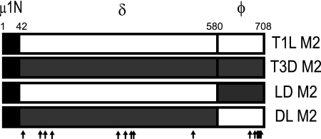
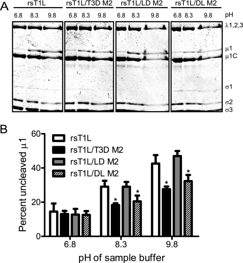
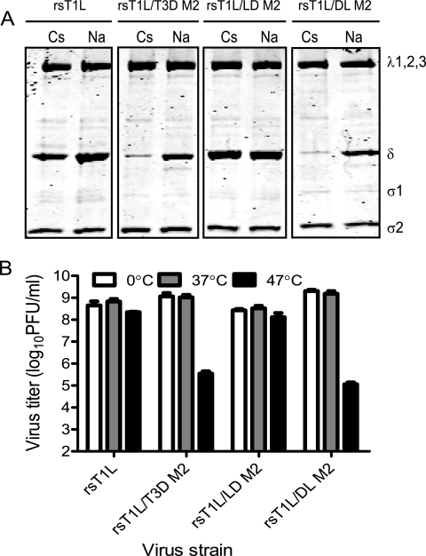
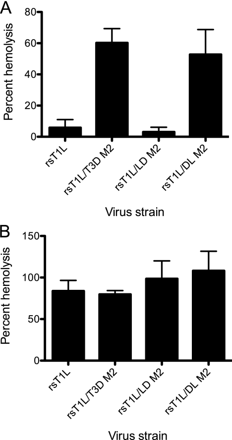
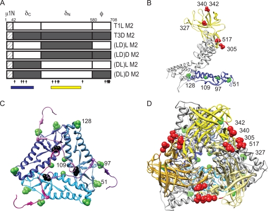
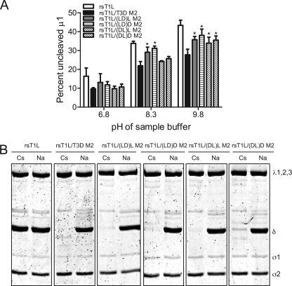
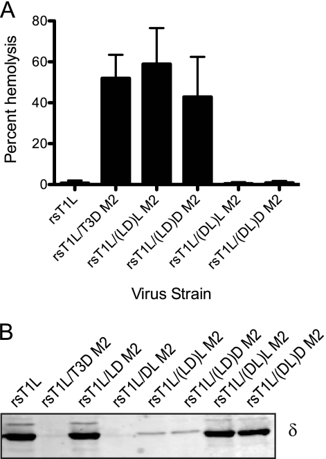
Cell entry of reovirus requires a series of ordered steps, which include conformational changes in outer capsid protein μ1 and its autocleavage. The μ1N fragment released as a consequence of these events interacts with host cell membranes and mediates their disruption, leading to delivery of the viral core into the cytoplasm. The prototype reovirus strains T1L and T3D exhibit differences in the efficiency of autocleavage, in the propensity to undergo conformational changes required for membrane penetration, and in the capacity for penetrating host cell membranes. To better understand how polymorphic differences in μ1 influence reovirus entry events, we generated recombinant viruses that express chimeric T1L-T3D μ1 proteins and characterized them for the capacity to efficiently complete each step required for membrane penetration. Our studies revealed two important functions for the central δ region of μ1. First, we found that μ1 autocleavage is regulated by the N-terminal portion of δ, which forms an α-helical pedestal structure. Second, we observed that the C-terminal portion of δ, which forms a jelly-roll β barrel structure, regulates membrane penetration by influencing the efficiency of ISVP* formation. Thus, our studies highlight the molecular basis for differences in the membrane penetration efficiency displayed by prototype reovirus strains and suggest that distinct portions of the reovirus δ domain influence different steps during entry.


Figures

FIG. 1.
FIG. 1.
Viruses with chimeric μ1 membrane penetration protein. Schematic diagrams of the μ1-encoding T1L and T3D M2 gene segments are shown, along with cleavage products generated during virus entry. Chimeric M2 gene segments (LD and DL) were constructed by reciprocally exchanging the δ- and φ-encoding portions of T1L and T3D M2 gene segments. The amino acid sequence of μ1N is identical for T1L and T3D and is shown as a hatched box. The locations of polymorphic residues in T1L and T3D μ1 are shown using vertical arrows.
FIG. 2.
FIG. 2.
The δ domain affects μ1N-μ1C cleavage efficiency. (A) CsCl-purified preparations of the indicated virus strains were mixed with reducing sample buffers at pH 6.8, 8.3, or 9.8. After incubation at 60°C for 5 min, the samples were resolved on SDS-10% PAGE gels and stained with Coomassie brilliant blue. (B) The intensities of the μ1 and μ1C bands were determined by densitometric analysis using a LI-COR Odyssey scanner. The fraction of uncleaved μ1 was determined by dividing the intensity of the μ1 band by the total intensity of the μ1 and μ1C bands. The results are expressed as the mean percent uncleaved μ1 quantified from three independent experiments. Error bars indicate the standard deviations (SD). *, P < 0.05, as determined by using the Student t test in comparison to rsT1L at the respective sample buffer pH.
FIG. 3.
FIG. 3.
The δ domain influences the capacity to undergo ISVP-to-ISVP* conversion. (A) ISVPs of the indicated viruses were treated with CsCl or NaCl at 30°C for 20 min, chilled on ice for 20 min, and treated with trypsin at 4°C for 30 min. Samples were resolved by SDS-PAGE and stained using Coomassie brilliant blue. The positions of reovirus capsid proteins are shown. (B) ISVPs of the indicated viruses were incubated at 37, or 47°C for 15 min. Residual infectivity was assessed by plaque assay. The results are shown as virus titers for triplicate samples. Error bars indicate the SD.
FIG. 4.
FIG. 4.
The δ domain modulates membrane penetration. A 3% (vol/vol) solution of bovine erythrocytes was incubated with 4.8 × 10¹⁰ ISVPs of the indicated viruses in virion-storage buffer containing no CsCl (A) or 300 mM CsCl (B) at 37°C for 1 h. Hemolysis was quantified by determining absorbance of the supernatant at 405 nm. Hemolysis after treatment of an equal number of cells with virion-storage buffer or virion-storage buffer containing 1% TX-100 was considered to be 0 or 100%, respectively. The results are expressed as the mean percent hemolysis for triplicate samples. Error bars indicate the SD.
FIG. 5.
FIG. 5.
Viruses with chimeric δ fragments. (A) Schematic diagram of the μ1-encoding T1L and T3D M2 gene segments are shown, along with cleavage products generated during virus entry. Chimeric M2 gene segments (LD)L, (LD)D, (DL)L, and (DL)D were constructed by reciprocally exchanging the δN- and δC-encoding portions of T1L and T3D M2 gene segments. The locations of polymorphic residues in T1L and T3D μ1 are indicated by vertical arrows. Regions spanning domain I and domain IV are indicated by blue and yellow boxes, respectively. (B) A side view of a μ1C monomer rendered using UCSF chimera from the crystal structure of μ1 (PDB:1JMU) is shown with domain I in blue and domain IV in yellow (38, 53). The positions of the polymorphic residues within domains I and IV are shown in green and red, respectively. Residue 97 is labeled since residue 96 was not resolved in the crystal structure and is not in the PDB file. (C) A close-up top view of the region near the autocleavage site is shown, with the domain I comprising δ in shades of blue and the μ1N fragment in shades of magenta. Polymorphic residues on one of the three chains are labeled as described for panel B. The autocleavage site is shown in black. (D) A top view of a μ1C trimer is shown, with domain I in shades of blue and domain IV in shades of yellow. Polymorphic residues on one of the three chains are labeled as described for panel B.
FIG. 6.
FIG. 6.
Different portions of δ modulate autocleavage efficiency and ISVP* formation. (A) CsCl-purified preparations of the indicated virus strains were mixed with reducing sample buffers at pH 6.8, 8.3, or 9.8. After incubation at 60°C for 5 min, the samples were resolved on SDS-10% PAGE gels and stained using Coomassie brilliant blue. The intensities of the μ1 and μ1C bands were determined by densitometric analysis using the LI-COR Odyssey scanner. The fraction of uncleaved μ1 was determined by dividing the intensity of the μ1 band by the total intensity of the μ1 and μ1C bands. The results are expressed as mean percent uncleaved μ1 for four independent experiments. Error bars indicate the SD. *, P < 0.05, as determined by using the Student t test in comparison to rsT1L/T3D M2 at the respective sample buffer pH. (B) ISVPs of the indicated viruses were treated with CsCl or NaCl at 30°C for 20 min, chilled on ice for 20 min, and treated with trypsin at 4°C for 30 min. Samples were resolved by SDS-PAGE and stained using Coomassie brilliant blue. The positions of reovirus capsid proteins are shown.
FIG. 7.
FIG. 7.
The jelly-roll β barrel domain affects efficiency of membrane penetration. (A) A 3% (vol/vol) solution of bovine erythrocytes was incubated with 4.8 × 10¹⁰ ISVPs of the indicated viruses in virion-storage buffer at 37°C for 1 h. Hemolysis was quantified by determining absorbance of the supernatant at 405 nm. Hemolysis after treatment of an equal number of cells with virion-storage buffer or virion-storage buffer containing 1% TX-100 was considered to be 0 or 100%, respectively. The results are expressed as mean percent hemolysis for six samples. Error bars indicate the SD. (B) A 3% (vol/vol) solution of bovine erythrocytes was incubated with 4.8 × 10¹⁰ ISVPs of the indicated viruses in virion-storage buffer at 37°C for 40 min, chilled on ice for 20 min, and treated with trypsin at 4°C for 30 min. Samples were resolved by SDS-PAGE and transferred to nitrocellulose membranes. The samples were probed with a μ1-specific MAb and visualized by using a LI-COR Odyssey scanner.

Similar articles

Cited by

References

    1. Agosto, M. A., T. Ivanovic, and M. L. Nibert. 2006. Mammalian reovirus, a nonfusogenic nonenveloped virus, forms size-selective pores in a model membrane. Proc. Natl. Acad. Sci. U. S. A. 103:16496-16501. - PMC - PubMed
    1. Arita, M., S. Koike, J. Aoki, H. Horie, and A. Nomoto. 1998. Interaction of poliovirus with its purified receptor and conformational alteration in the virion. J. Virol. 72:3578-3586. - PMC - PubMed
    1. Baer, G. S., and T. S. Dermody. 1997. Mutations in reovirus outer-capsid protein σ3 selected during persistent infections of L cells confer resistance to protease inhibitor E64. J. Virol. 71:4921-4928. - PMC - PubMed
    1. Banerjee, M., and J. E. Johnson. 2008. Activation, exposure and penetration of virally encoded, membrane-active polypeptides during non-enveloped virus entry. Curr. Protein Peptide Sci. 9:16-27. - PubMed
    1. Barton, E. S., J. L. Connolly, J. C. Forrest, J. D. Chappell, and T. S. Dermody. 2001. Utilization of sialic acid as a coreceptor enhances reovirus attachment by multistep adhesion strengthening. J. Biol. Chem. 276:2200-2211. - PubMed

Publication types

LinkOut - more resources