. 2010 Dec;84(24):12609-18.
doi: 10.1128/JVI.01400-10. Epub 2010 Oct 13.

The matrix protein of vesicular stomatitis virus binds dynamin for efficient viral assembly

Affiliations

The matrix protein of vesicular stomatitis virus binds dynamin for efficient viral assembly

Hélène Raux et al. J Virol. 2010 Dec.

Abstract

Matrix proteins (M) direct the process of assembly and budding of viruses belonging to the Mononegavirales order. Using the two-hybrid system, the amino-terminal part of vesicular stomatitis virus (VSV) M was shown to interact with dynamin pleckstrin homology domain. This interaction was confirmed by coimmunoprecipitation of both proteins in cells transfected by a plasmid encoding a c-myc-tagged dynamin and infected by VSV. A role for dynamin in the viral cycle (in addition to its role in virion endocytosis) was suggested by the fact that a late stage of the viral cycle was sensitive to dynasore. By alanine scanning, we identified a single mutation of M protein that abolished this interaction and reduced virus yield. The adaptation of mutant virus (M.L4A) occurred rapidly, allowing the isolation of revertants, among which the M protein, despite having an amino acid sequence distinct from that of the wild type, recovered a significant level of interaction with dynamin. This proved that the mutant phenotype was due to the loss of interaction between M and dynamin. The infectious cycle of the mutant virus M.L4A was blocked at a late stage, resulting in a quasi-absence of bullet-shaped viruses in the process of budding at the cell membrane. This was associated with an accumulation of nucleocapsids at the periphery of the cell and a different pattern of VSV glycoprotein localization. Finally, we showed that M-dynamin interaction affects clathrin-dependent endocytosis. Our study suggests that hijacking the endocytic pathway might be an important feature for enveloped virus assembly and budding at the plasma membrane.


Figures

FIG. 1.
FIG. 1.
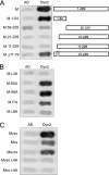
VSV M protein binds dynamin. (A) Mapping studies of M protein binding domain within dynamin 2. The interactions between M protein fused to the DNA-binding domain of LexA and different fragments of dynamin fused to Gal4 activation domain (AD) were assessed by the appearance of blue colonies in the presence of X-Gal (5-bromo-4-chloro-3-indolyl-β-d-galactopyranoside). The fragment of dynamin 2 extending from aa 528 to 828 corresponds to the clone isolated during the screen. (B) Detection of M-dynamin complex in cells. (A) HEK293T cells were transfected (+) or not (−) with plasmid pDyn2/c-Myc. At 24 h posttransfection, cells were mock infected (NI) or infected (VSV) by wild-type VSV at an MOI of 5 PFU/cell. At 4 h after infection, cells were harvested and lysed. Cell lysate was incubated with a mouse polyclonal anti-Mvsv antibody. Immune complexes were precipitated by incubation with protein A-Sepharose. Immunoprecipitated proteins (IP) were analyzed by Western immunoblotting with mouse monoclonal anti-dynamin antibody. In the left portion of the blot, direct cellular extracts (C) of transfected cells (either mock infected [NI] or infected by VSV) were also analyzed.
FIG. 2.
FIG. 2.
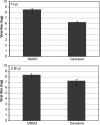
Dynasore inhibits VSV viral cycle at a late stage. Monolayers of BSR cells were infected with WT VSV at an MOI of 3 PFU/cell. After 1 h or 2.5 h of infection, the medium was changed for DMEM containing 80 μM dynasore. The culture supernatants were collected at 5 h postinfection. Viral production was determined by a plaque assay. The results are the average of three independent experiments.
FIG. 3.
FIG. 3.
Mapping studies of the dynamin 2 binding domain within M protein and the interaction of dynamin 2 with the M proteins of other vesiculoviruses. (A) The interactions between deletion mutants of M protein fused to the DNA-binding domain of LexA and the fragment (aa 528 to 828) of dynamin 2 fused to Gal4 AD were assayed as in Fig. 1A. (B) Refinement of the analysis of dynamin 2 binding domain within M protein by alanine scanning. The interactions between mutant M proteins (in which amino acids 4 to 8 have been replaced by an alanine) fused to the DNA-binding domain of LexA and the fragment (aa 528 to 828) of dynamin 2 fused to Gal4 AD were assayed as in Fig. 1A. (C) Conservation of the interaction between M and dynamin 2 among the vesiculovirus genus. Interactions between CV and SVCV M proteins and the C-terminal part of dynamin 2 (residues 528 to 828) were assayed as in Fig. 1A. Since the full-length M protein of SVCV fused to the DNA-binding domain of LexA activated the expression of β-galactosidase when expressed alone, we could only assess the interaction between the amino-terminal part of SVCV M protein (residues 1 to 34) and dynamin. The interaction of mutant L4A of CV M protein with dynamin was also analyzed.
FIG. 4.
FIG. 4.
Characterization of the production of recombinant virus M.L4A and its revertants. (A) Protein profiles for WT VSV, M.L4A recombinant virus, and its revertants rM.S2I-L4A, rM.S2R-L4A, rM.S3F-L4A, and rM.L4V. Virions were harvested from supernatant of infected BSR cells at 6 h postinfection. Virion proteins were analyzed by SDS-PAGE and Coomassie blue staining. The percent yield of each protein in this experiment was calculated using ImageJ software on a scan of the gel. (B) Lysates of the infected BSR cells were analyzed by Western blotting with an anti-VSV polyclonal serum and a monoclonal antibody directed against tubulin. (C) Growth kinetics of WT VSV (⋄), M.L4A recombinant virus (▵), and its revertant rM.S3F-L4A (○). BSR cells were infected at an MOI of 0.3 (left side graph) or 3 (right side graph), and samples were harvested for titration at the indicated times postinfection. Virus titers represent averages from at least three independent experiments. The y scale is not the same on both graphs.
FIG. 5.
FIG. 5.
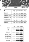
Characterization of revertant phenotypes. (A) Comparison of the morphology of the plaques of WT VSV, M.L4A recombinant virus, and revertant rM.S3F-L4A. (B) Nucleotide sequence of the 5′ end of the open reading frame of M mRNA for wild-type, mutant, and revertant viruses. (C) Characterization of the binding between M proteins (WT, L4A, S2I-L4A, S2R-L4A, S3F-L4A, and L4V) and the C-terminal fragments of dynamins 1 (aa 523 to 850) and 2 (aa 528 to 828) using the yeast two-hybrid system.
FIG. 6.
FIG. 6.
Electron micrographs of BSR cells infected by WT VSV, M.L4A recombinant virus, and rM.S3F-L4A revertant at 5 h postinfection. Size bars, 500 nm. Virions in the process of budding at the plasma membrane of cells infected by the different viruses are indicated by arrowheads.
FIG. 7.
FIG. 7.
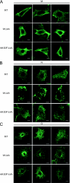
Localization of M, N, and G proteins in cells infected by WT VSV, mutant M.L4A, or revertant rM.S3F-L4A. BSR cells were infected by WT VSV, mutant M.L4A, or revertant rM.S3F-L4A. At 4 h postinfection, the cells were analyzed by immunofluorescence with specific antibodies to detect M protein (A), N protein (B), and G protein (C); the staining patterns of M, N, and G are shown for different cells. Note the accumulation of N near the plasma membrane for mutant M.L4A. Size bars, 10 μm.
FIG. 8.
FIG. 8.
WT VSV M protein affects clathrin-dependent endocytosis. (A and B) Tfn endocytosis in cells infected by WT or M.L4A viruses. Cells were infected at an MOI of 0.4 (A) or 3 (B). At 3.5 h postinfection, Alexa 488-conjugated Tfn (green) was added to the medium for 25 min. Cells were washed and fixed. In panel A, after fixation, infected cells were labeled with monoclonal anti-N antibody (red) and, for WT virus, uninfected cells are indicated by arrowheads. In panel B, noninfected cells (NI) are shown as a positive control of endocytosis. Arrowheads indicate cells infected by WT virus with fluorescent Tfn-containing vesicles accumulated just beneath the plasma membrane. Size bars, 32 μm.

Similar articles

Cited by

References

    1. Bergmann, J. E., and P. J. Fusco. 1988. The M protein of vesicular stomatitis virus associates specifically with the basolateral membranes of polarized epithelial cells independently of the G protein. J. Cell Biol. 107:1707-1715. - PMC - PubMed
    1. Bourhis, J. M., B. Canard, and S. Longhi. 2006. Structural disorder within the replicative complex of measles virus: functional implications. Virology 344:94-110. - PubMed
    1. Brown, E. L., and D. S. Lyles. 2003. Organization of the vesicular stomatitis virus glycoprotein into membrane microdomains occurs independently of intracellular viral components. J. Virol. 77:3985-3992. - PMC - PubMed
    1. Bruce, E. A., P. Digard, and A. D. Stuart. 2010. The Rab11 pathway is required for influenza A virus budding and filament formation. J. Virol. 84:5848-5859. - PMC - PubMed
    1. Cao, H., F. Garcia, and M. A. McNiven. 1998. Differential distribution of dynamin isoforms in mammalian cells. Mol. Biol. Cell 9:2595-2609. - PMC - PubMed

Publication types

LinkOut - more resources