. 2010 Nov 2;107(44):18997-9001.
doi: 10.1073/pnas.1004380107. Epub 2010 Oct 13.

Inhibition of pathogen-induced apoptosis by a Coxiella burnetii type IV effector protein

Affiliations

Inhibition of pathogen-induced apoptosis by a Coxiella burnetii type IV effector protein

Anja Lührmann et al. Proc Natl Acad Sci U S A. .

Abstract

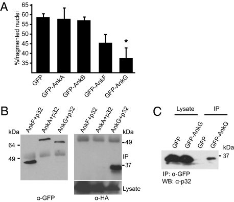
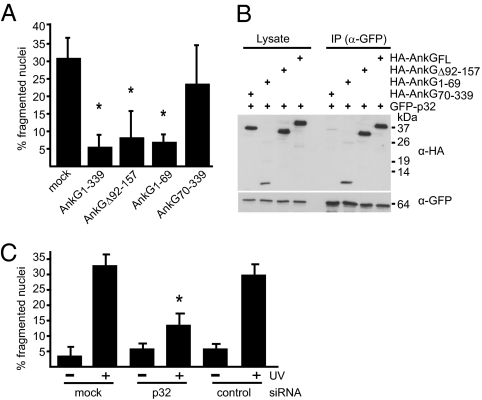
Coxiella burnetii and Legionella pneumophila are evolutionarily related pathogens with different intracellular infection strategies. C. burnetii persists within and is transmitted by mammalian hosts, whereas, L. pneumophila is found primarily in the environment associated with protozoan hosts. Although a type IV secretion system encoded by the defect in organelle trafficking (dot) and intracellular multiplication (icm) genes is a virulence determinant that remains highly conserved in both bacteria, the two pathogens encode a different array of effector proteins that are delivered into host cells by the Dot/Icm machinery. This difference suggests that adaptations to evolutionarily distinct hosts may be reflected in the effector protein repertoires displayed by these two pathogens. Here we provide evidence in support of this hypothesis. We show that a unique C. burnetii effector from the ankyrin repeat (Ank) family called AnkG interferes with the mammalian apoptosis pathway. AnkG was found to interact with the host protein gC1qR (p32). Either the addition of AnkG to the repertoire of L. pneumophila effector proteins or the silencing of p32 in mouse dendritic cells resulted in a gain of function that allowed intracellular replication of L. pneumophila in these normally restrictive mammalian host cells by preventing rapid pathogen-induced apoptosis. These data indicate that p32 regulates pathogen-induced apoptosis and that AnkG functions to block this pathway. Thus, emergence of an effector protein that interferes with a proapoptotic signaling pathway directed against intracellular bacteria correlates with adaptation of a pathogen to mammalian hosts.


Conflict of interest statement

The authors declare no conflict of interest.

Figures

Fig. 1.
Fig. 1.
AnkG interferes with apoptosis and binds the host protein p32. (A) CHO cells ectopically expressing GFP or the indicated GFP-tagged C. burnetii Anks were incubated with staurosporine. The nuclei were stained with DAPI, and nuclei of GFP-expressing cells were scored for fragmentation. One hundred nuclei from GFP-expressing cells per sample from four independent experiments were counted. *P < 0.001. (B) HEK 293 cells were cotransfected with plasmids encoding HA-tagged p32 and the indicated GFP-tagged C. burnetii ankyrin repeat-containing proteins. Proteins were precipitated from the cell lysates with an anti-GFP antibody and analyzed by immunoblot. The α-GFP blot shows Ank protein levels in the immunoprecipitate (IP), and the α-HA blot shows p32 levels in the immunoprecipitate (IP) and lysate. (C) HEK 293 cells were transfected with a plasmid encoding GFP-AnkG or the GFP control. Proteins were precipitated from the cell lysates with an anti-GFP antibody, and endogenous p32 was analyzed by immunoblotting with an anti-p32 antibody.
Fig. 2.
Fig. 2.
The AnkG antiapoptotic domain mediates binding to the proapoptotic protein p32. (A) CHO cells ectopically expressing the indicated HA-tagged truncations of AnkG were incubated with staurosporine. The cells were stained with DAPI and HA-specific antibody. The nuclear morphology of HA-expressing cells was scored as in Fig.1A. *P < 0.003. (B) HEK 293 cells were cotransfected with plasmids encoding GFP-tagged p32 and the indicated HA-tagged truncations of AnkG. Proteins were precipitated from the cell lysates with an anti-GFP antibody. Immunoblot analysis was used to detect p32 (α-GFP) and AnkG (α-HA) in the lysates and immunoprecipitates (IP). (C) HeLa cells transfected with control siRNA (control), with p32 siRNA (p32), or untransfected (mock) were exposed to UV light as indicated. The cells were stained with DAPI, and nuclear morphology was scored. The percentage of cells with apoptotic nuclear morphology was calculated. One hundred nuclei per sample from three independent experiments were counted. *P < 0.006.
Fig. 3.
Fig. 3.
Translocation of AnkG prevents rapid pathogen-induced apoptosis, allowing L. pneumophila replication in DCs. (A) CHO-FcR cells infected with L. pneumophila harboring the vector pJV400, pJV400-AnkB (AnkB), or pJV400-AnkG (AnkG) were incubated with staurosporine. L. pneumophila was stained with a specific anti-Legionella antibody, and the nuclei were scored by the TUNEL assay. The nuclei of at least 100 infected cells per sample from three independent experiments were counted. *P < 0.01. (B) Bone marrow DCs derived from B6 mice were infected for 6 h with L. pneumophila ΔflaA expressing the pJV400 vector, AnkG, or AnkB or with L. pneumophila ΔdotA expressing AnkG. L. pneumophila was stained with a specific anti-Legionella antibody, and the nuclei of infected cells were scored by the TUNEL assay. Data shown are the mean ± SD of 300 cells counted per each coverslip in triplicate and are representative of two independent experiments. **P < 0.01. (C and D) Representative fluorescence micrographs of B6 DCs infected with (C) L. pneumophila ΔflaA + pJV400, ΔflaA + AnkB, or ΔflaA + AnkG and (D) L. pneumophila ΔflaA expressing AnkG1–69 or AnkG70–339 or L. pneumophila ΔdotA expressing full-length AnkG. DCs were fixed at 10 h postinfection and were stained with an antibody specific for MHC class II (red), DAPI (blue), and an anti-Legionella antibody (green).
Fig. 4.
Fig. 4.
Rapid pathogen-induced apoptosis in DCs requires p32 function. (A) DCs untreated, treated with siRNA against p32, or mock treated were infected with L. pneumophila ΔflaA, ΔflaA expressing AnkG, or ΔdotA. L. pneumophila were stained with a specific anti-Legionella antibody, and the nuclei of infected cells were scored using DAPI staining. Data shown are from one experiment representative of five independent experiments that yielded similar results. n = 200 MHC class II-positive DCs. (B) DCs untreated (black bars), treated with siRNA against p32 (white bars), or mock treated (gray bars) were infected with L. pneumophila ΔflaA, ΔflaA expressing AnkG, or ΔdotA. Vacuoles containing replicating bacteria at 10 h postinfection were counted. Data shown are from one representative experiment representative of five independent experiments that yielded similar results. n = 200 MHC class II-positive DCs. N.D., vacuoles containing replicating bacteria were not detectable; R.V., vacuoles containing replicating bacteria. (C) DCs treated with p32 siRNA (Right) or mock treated (Left) were infected with L. pneumophila ΔflaA or with L. pneumophila WT. Vacuoles containing replicating bacteria at 10 h postinfection were counted. Data are shown from one experiment representative of two independent experiments that yielded similar results. n = 200 MHC class II-positive DCs.

References

    1. Fields BS. The molecular ecology of Legionellae. Trends Microbiol. 1996;4:286–290. - PubMed
    1. Ninio S, Roy CR. Effector proteins translocated by Legionella pneumophila: Strength in numbers. Trends Microbiol. 2007;15:372–380. - PubMed
    1. Ensminger AW, Isberg RR. Legionella pneumophila Dot/Icm translocated substrates: A sum of parts. Curr Opin Microbiol. 2009;12:67–73. - PMC - PubMed
    1. Isberg RR, O'Connor TJ, Heidtman M. The Legionella pneumophila replication vacuole: Making a cosy niche inside host cells. Nat Rev Microbiol. 2009;7:13–24. - PMC - PubMed
    1. Fraser DW, et al. Legionnaires’ disease: Description of an epidemic of pneumonia. N Engl J Med. 1977;297:1189–1197. - PubMed

Publication types

MeSH terms