. 2010 Oct 1;66(Pt 10):1160-6.
doi: 10.1107/S1744309110002514. Epub 2010 Mar 5.

The structure of SSO2064, the first representative of Pfam family PF01796, reveals a novel two-domain zinc-ribbon OB-fold architecture with a potential acyl-CoA-binding role

Affiliations

The structure of SSO2064, the first representative of Pfam family PF01796, reveals a novel two-domain zinc-ribbon OB-fold architecture with a potential acyl-CoA-binding role

S Sri Krishna et al. Acta Crystallogr Sect F Struct Biol Cryst Commun. .

Abstract

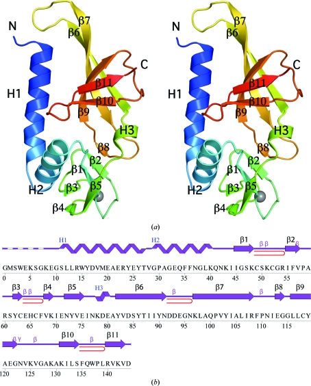
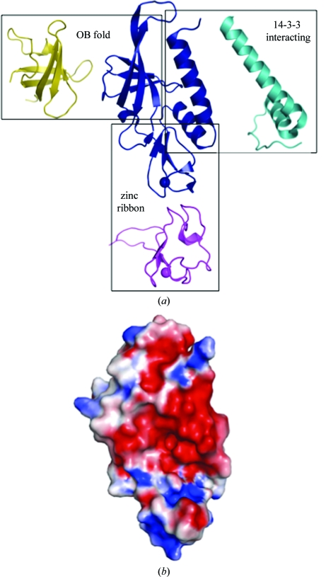
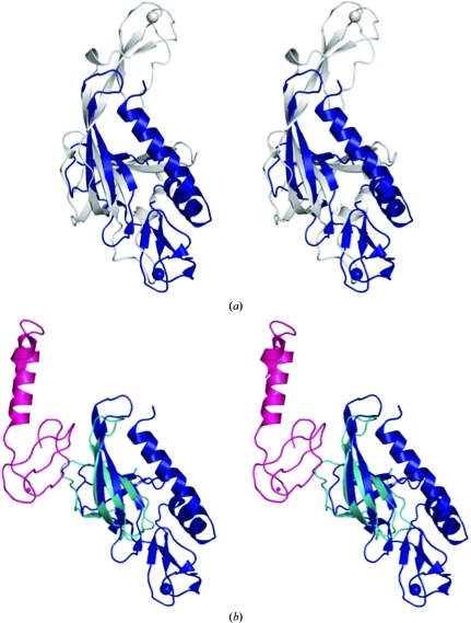
SSO2064 is the first structural representative of PF01796 (DUF35), a large prokaryotic family with a wide phylogenetic distribution. The structure reveals a novel two-domain architecture comprising an N-terminal, rubredoxin-like, zinc ribbon and a C-terminal, oligonucleotide/oligosaccharide-binding (OB) fold domain. Additional N-terminal helical segments may be involved in protein-protein interactions. Domain architectures, genomic context analysis and functional evidence from certain bacterial representatives of this family suggest that these proteins form a novel fatty-acid-binding component that is involved in the biosynthesis of lipids and polyketide antibiotics and that they possibly function as acyl-CoA-binding proteins. This structure has led to a re-evaluation of the DUF35 family, which has now been split into two entries in the latest Pfam release (v.24.0).


Figures

Figure 1
Figure 1
Crystal structure of SSO2064 from S. solfataricus. (a) Stereo ribbon diagram of the SSO2064 monomer (chain A) color-coded from the N-terminus (blue) to the C-terminus (red). Helices (H1–H3) and β-strands (β1–β11) are indicated. The zinc ion is depicted as a gray sphere. (b) Diagram showing the secondary-structure elements of SSO2064 superimposed on its primary sequence. The labeling of secondary-structure elements is in accord with PDBsum (http://www.ebi.ac.uk/pdbsum): α-helices are labeled H1 and H2, the 310-helix is labeled H3, the β-strands are labeled β1–β11, β-turns and γ-turns are designated by their respective Greek letters (β, γ) and red loops indicate β-hairpins.
Figure 2
Figure 2
SSO2064 exhibits structural similarity to protein–protein interaction motifs, rubredoxin-like zinc ribbons and OB folds. (a) Ribbon diagram showing the structural superposition of SSO2064 (PDB code 3irb; residues 8–144; blue) with the 14-3-3 interacting motif of plant H+-ATPase (PDB code 2o98; residues 7–56; cyan), a zinc-substituted rubredoxin from Guillardia theta (PDB code 1h7v; residues 1–60; magenta) and the major cold-shock protein from E. coli (PDB code 1mjc; residues 1–70; yellow). Superposed proteins have been translated for clarity. Zinc ions are indicated as spheres. (b) Electrostatic surface representation of SSO2064 in the same orientation as in (a). Positive potential is in blue (+7kTe−1) and negative potential is in red (−7kTe−1).
Figure 3
Figure 3
SSO2064 subdomains display a novel spatial organization with respect to other OB-fold-containing and zinc-ribbon-containing proteins. Stereo ribbon diagram showing the structural superposition of SSO2064 (PDB code 3irb; residues 8–144; blue) with (a) the mini-chromosome maintenance complex (MCM) from M. thermoautotrophicum (PDB code 1ltl; residues 95–242; gray) and (b) chains A (residues 28–83; cyan) and Y (residues 10–71; magenta) from the large ribosomal subunit from H. marismortui (PDB code 1jj2; Klein et al., 2001 ▶). Zinc (gray and blue) and cobalt (magenta) [chain Y in (b)] are indicated as spheres.

References

    1. Anantharaman, V., Koonin, E. V. & Aravind, L. (2001). J. Mol. Biol.307, 1271–1292. - PubMed
    1. Arcus, V. (2002). Curr. Opin. Struct. Biol.12, 794–801. - PubMed
    1. Baker, N. A., Sept, D., Joseph, S., Holst, M. J. & McCammon, J. A. (2001). Proc. Natl Acad. Sci. USA, 98, 10037–10041. - PMC - PubMed
    1. Bangera, M. G. & Thomashow, L. S. (1999). J. Bacteriol.181, 3155–3163. - PMC - PubMed
    1. Berman, H. M. et al. (2009). Nucleic Acids Res.37, D365–D368. - PMC - PubMed

Publication types

Associated data