. 2010 Oct 1;66(Pt 10):1167-73.
doi: 10.1107/S1744309110007517. Epub 2010 Jul 6.

Structure of the first representative of Pfam family PF04016 (DUF364) reveals enolase and Rossmann-like folds that combine to form a unique active site with a possible role in heavy-metal chelation

Affiliations

Structure of the first representative of Pfam family PF04016 (DUF364) reveals enolase and Rossmann-like folds that combine to form a unique active site with a possible role in heavy-metal chelation

Mitchell D Miller et al. Acta Crystallogr Sect F Struct Biol Cryst Commun. .

Abstract

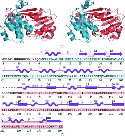
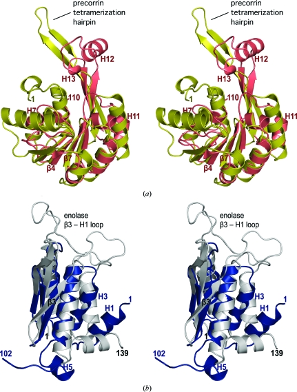
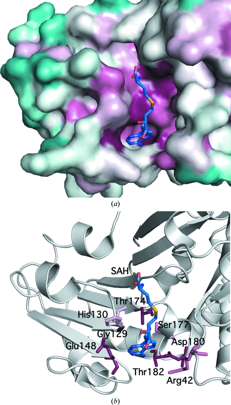
The crystal structure of Dhaf4260 from Desulfitobacterium hafniense DCB-2 was determined by single-wavelength anomalous diffraction (SAD) to a resolution of 2.01 Å using the semi-automated high-throughput pipeline of the Joint Center for Structural Genomics (JCSG) as part of the NIGMS Protein Structure Initiative (PSI). This protein structure is the first representative of the PF04016 (DUF364) Pfam family and reveals a novel combination of two well known domains (an enolase N-terminal-like fold followed by a Rossmann-like domain). Structural and bioinformatic analyses reveal partial similarities to Rossmann-like methyltransferases, with residues from the enolase-like fold combining to form a unique active site that is likely to be involved in the condensation or hydrolysis of molecules implicated in the synthesis of flavins, pterins or other siderophores. The genome context of Dhaf4260 and homologs additionally supports a role in heavy-metal chelation.


Figures

Figure 1
Figure 1
Crystal structure of Dhaf4260 from D. hafniense. (a) Stereo ribbon diagram of the Dhaf4260 monomer. The N-terminal domain is colored cyan and the C-terminal domain is colored salmon. Helices H1–H13 and β-strands β1–β10 are indicated. (b) Diagram showing the secondary-structure elements of Dhaf4260 superimposed on its primary sequence. The designation of secondary-structure elements is in accord with PDBsum (http://www.ebi.ac.uk/pdbsum). For Dhaf4260, helices are labeled sequentially (H1, H2, H3 etc.) with α-helices H1–H6, H8–H10 and H12 and 310-helices H7, H11 and H13, β-strands are numbered sequentially (strands β1–β3 form the first sheet and strands β4–β10 form the second sheet), β-turns are labeled β, γ-turns are labeled γ and β-hairpins are indicated as red loops. The unmodeled sequence, which is disordered in the electron-density map, is indicated by a dashed line. Residues from the N-terminal domain are highlighted in cyan and residues from the C-terminal domain are in salmon.
Figure 2
Figure 2
Stereo ribbon diagram showing the structural superposition of (a) the C-terminal domain of Dhaf4260 (PDB code 3l5o; residues 110–251; salmon) and precorrin-8w methyltransferase from Methanobacterium thermoautotrophicum (MT0146; PDB code 1f38; residues 1–186; gold) and (b) the N-terminal domain of Dhaf4260 (PDB code 3l5o; residues 1–102; blue) and the enolase N-terminal domain from Saccharomyces cerevisiae (PDB code 4enl; residues 1–139; gray). The precorrin methyltransferase and enolase regions implicated in oligomerization and substrate binding are indicated.
Figure 3
Figure 3
The interdomain pocket forms a unique catalytic site. (a) Surface representation of the Dhaf4260 domain interface colored by sequence conservation according to ConSurf (Landau et al., 2005 ▶). High conservation among DUF364 homologs is indicated in maroon and low conservation is indicated in turquoise. A docked S-­adenosyl-l-homocysteine (SAH) molecule is shown in ball-and-stick representation. Docking was based on its superposition with MT0146 (PDB code 1l3i; Keller et al., 2002 ▶). (b) Ribbon representation of Dhaf4260 in the same orientation as in (a). Highly conserved Dhaf4260 residues are shown in ball-and-stick representation and are labeled.

References

    1. Burroughs, A. M., Allen, K. N., Dunaway-Mariano, D. & Aravind, L. (2006). J. Mol. Biol.361, 1003–1034. - PubMed
    1. Carugo, O. & Argos, P. (1997). Proteins, 28, 10–28. - PubMed
    1. Cohen, A. E., Ellis, P. J., Miller, M. D., Deacon, A. M. & Phizackerley, R. P. (2002). J. Appl. Cryst.35, 720–726. - PMC - PubMed
    1. Collaborative Computational Project, Number 4 (1994). Acta Cryst. D50, 760–763. - PubMed
    1. Cruickshank, D. W. J. (1999). Acta Cryst. D55, 583–601. - PubMed

Publication types

Associated data