

Review
doi: 10.1107/S1744309110007177.
Epub 2010 Jul 6.
Structural classification of proteins and structural genomics: new insights into protein folding and evolution
Affiliations
- PMID: 20944210
- PMCID: PMC2954204
- DOI: 10.1107/S1744309110007177
Item in Clipboard
Review
Structural classification of proteins and structural genomics: new insights into protein folding and evolution
Acta Crystallogr Sect F Struct Biol Cryst Commun.
.
Abstract
During the past decade, the Protein Structure Initiative (PSI) centres have become major contributors of new families, superfamilies and folds to the Structural Classification of Proteins (SCOP) database. The PSI results have increased the diversity of protein structural space and accelerated our understanding of it. This review article surveys a selection of protein structures determined by the Joint Center for Structural Genomics (JCSG). It presents previously undescribed β-sheet architectures such as the double barrel and spiral β-roll and discusses new examples of unusual topologies and peculiar structural features observed in proteins characterized by the JCSG and other Structural Genomics centres.
Figures

Gallery of selected protein structures determined by the JCSG (see also Figs. 5 ▶, 6 ▶ and 7 ▶). (a) Acetoacetate decarboxylase (ADC) subunit (PBD entry 3c8w ). β-Strands in the double-barrel β-sheet are shown as coloured arrows; other secondary-structure elements and loops are shown as silver coils. (b) DUF1089 protein PA1994 (PDB entry 2h1t ) coloured by rainbow. An ‘unswapped’ monomer is shown, a large β-sheet of which is folded into a spiral roll. (c) DUF1831 protein lp2179 (PDB entry 2iay ) coloured by secondary structure: red, α-helix; yellow, β-strand; green, loop. (d) DUF1470 protein Jann2411 (PDB entry 3h0n ) coloured by secondary structure, with the N-terminal subdomain coloured as in (c) and the C-terminal subdomain coloured using an alternative palette: cyan, α-helix; purple, β-strand; pink, loop. The sphere represents the zinc ion. (e) DUF1488 protein Shew3726 (PDB entry 2gpi ) coloured by secondary structure, with the rare left-handed β-X-β unit coloured using an alternative palette. (f) DUF2006 protein NE1406 (PDB entry 2ich ) viewed along the pseudo-twofold axis that relates its similar barrel domains. The topologically equivalent β-strands in both domains and in ADC (a) are shown in the same colour.

New β-sheet architectures. (a) A bifurcated X-shaped β-sheet can fold upon itself on both sides, forming a double barrel. (b) A very large β-sheet can be folded into a β-spiral roll with overlapping edges. This architecture combines features of both β-barrel and β-sandwich. In both parts, for simplicity, the arrows denote β-strands but do not define the strand directionality. The actual β-sheets of these architectures may comprise parallel and antiparallel strands.

A trefoil knot in the structure of the uncharacterized protein MJ0366 (a duplicated RHH motif).

(a) The structure of UPF0352 protein CPS2611 (PDB entry 2ota ; S. M. Vorobiev, W. Zhou, M. Su, J. Seetharaman, H. Wang, H. Janjua, K. Cunningham, L.-C. Ma, C. Liu, T. B. Acton, R. Xiao, G. T. Montelione, L. Tong & J. F. Hunt, unpublished work) is an example of an obligatory oligomer. It is composed of two interlocking noncompact subunits, which are coloured orange and blue. (b) A representative structure of the DinB-like family member (PDB entry 2f22 ; JCSG, unpublished work). It contains two interlocking structural repeats, which are shown in green and red.

Structures of the CsrA dimer (a) and the C-terminal domain of YqeH (b). The individual subunits of CsrA are coloured cyan and blue, whereas the YqeH structural repeats are shown in yellow and red. (c) Stereoview of the superimposition of the CsrA dimer (blue) and the C-terminal domain of YqeH (orange).
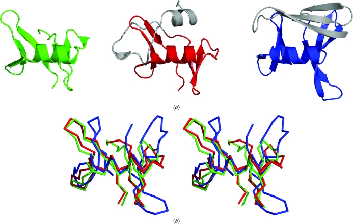
A consensus fold of the N- and C-terminal domains of the DUF1285 family. (a) Side-by-side comparison of the N-terminal domains of 2ra9 (green) and 2re3 (red) and the C-terminal domain of 2re3 (blue). Nonconserved additional regions are shown in grey. (b) Stereoview of the superimposition of the common parts of the three domains.
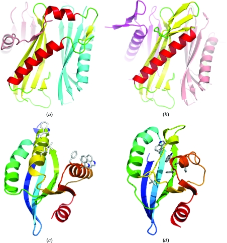
Metamorphic proteins. (a, b)Side-by-side comparison of alternatively folded subunits of the DUF74 pentamer (PDB entry 1vr4 ). Chain A and chain D (b) are coloured according to their secondary structure. The adjacent subunits are coloured as follows: chain B, light blue; chain C, magenta; chain E, pink. (c, d) Side-by-side comparison of the Sfri0576-like family structures 2q3l (c) and 2ook (d). The equivalent nonpolar residues that are exposed on the 2q3l surface and buried in the 2ook core are shown in stick representation.
References
Publication types
MeSH terms
Substances
Grants and funding
LinkOut - more resources
Full Text Sources
Miscellaneous
