. 2010 Oct 1;66(Pt 10):1274-80.
doi: 10.1107/S1744309110032999. Epub 2010 Sep 22.

Structure of BT_3984, a member of the SusD/RagB family of nutrient-binding molecules

Affiliations

Structure of BT_3984, a member of the SusD/RagB family of nutrient-binding molecules

Constantina Bakolitsa et al. Acta Crystallogr Sect F Struct Biol Cryst Commun. .

Abstract

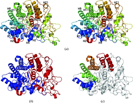
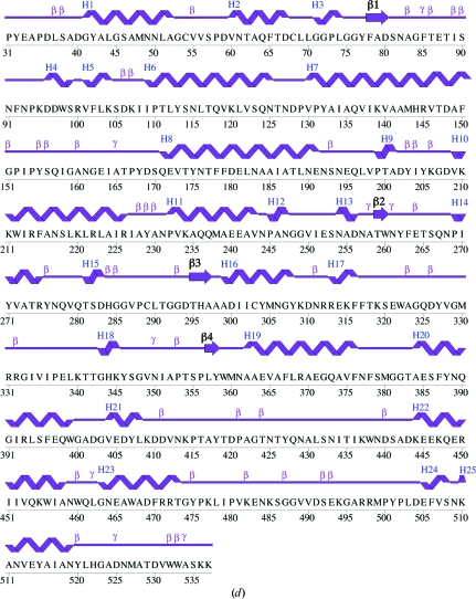
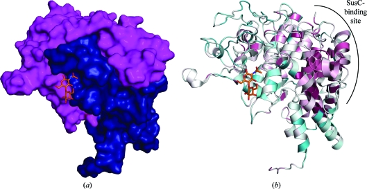
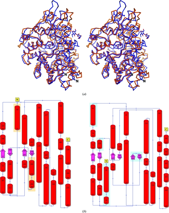
The crystal structure of the Bacteroides thetaiotaomicron protein BT_3984 was determined to a resolution of 1.7 Å and was the first structure to be determined from the extensive SusD family of polysaccharide-binding proteins. SusD is an essential component of the sus operon that defines the paradigm for glycan utilization in dominant members of the human gut microbiota. Structural analysis of BT_3984 revealed an N-terminal region containing several tetratricopeptide repeats (TPRs), while the signature C-terminal region is less structured and contains extensive loop regions. Sequence and structure analysis of BT_3984 suggests the presence of binding interfaces for other proteins from the polysaccharide-utilization complex.


Figures

Figure 1
Figure 1
Crystal structure of BT_3984 from B. thetaiotaomicron VPI-5482. (a) Stereo ribbon diagram of the BT_3984 monomer color-coded from the N-terminus (blue) to the C-­terminus (red). Helices H1–H25 and β-strands (β1–β4) are indicated. (b) Ribbon diagram in the same orientation as (a) showing the two subdomains of BT-3984 colored in blue and red for the N- and C-terminal regions, respectively. (c) Ribbon diagram in the same orientation as in (a) showing the four TPRs present in BT_3984: from the N- to C-terminus, TPR1 (blue), TPR2 (green), TPR3 (orange) and TPR4 (red). (d) Diagram showing the secondary-structure elements of BT_3984 superimposed on its primary sequence. The labeling of secondary-structure elements is in accord with PDBsum (http://www.ebi.ac.uk/pdbsum), where α-helices are labeled H1, H2, H3 etc., β-strands are labeled and β-turns and γ-turns are designated by their respective Greek letters (β, γ). For BT_3984, the α-helices (H1–H8, H10–H11, H14, H16–H17 and H19–H25), 310-­helices (H9, H12–H13, H15 and H18) and β-strands (β1–4) are indicated.
Figure 1
Figure 1
Crystal structure of BT_3984 from B. thetaiotaomicron VPI-5482. (a) Stereo ribbon diagram of the BT_3984 monomer color-coded from the N-terminus (blue) to the C-­terminus (red). Helices H1–H25 and β-strands (β1–β4) are indicated. (b) Ribbon diagram in the same orientation as (a) showing the two subdomains of BT-3984 colored in blue and red for the N- and C-terminal regions, respectively. (c) Ribbon diagram in the same orientation as in (a) showing the four TPRs present in BT_3984: from the N- to C-terminus, TPR1 (blue), TPR2 (green), TPR3 (orange) and TPR4 (red). (d) Diagram showing the secondary-structure elements of BT_3984 superimposed on its primary sequence. The labeling of secondary-structure elements is in accord with PDBsum (http://www.ebi.ac.uk/pdbsum), where α-helices are labeled H1, H2, H3 etc., β-strands are labeled and β-turns and γ-turns are designated by their respective Greek letters (β, γ). For BT_3984, the α-helices (H1–H8, H10–H11, H14, H16–H17 and H19–H25), 310-­helices (H9, H12–H13, H15 and H18) and β-strands (β1–4) are indicated.
Figure 2
Figure 2
Structural organization of BT_3984 and homologs. (a) Surface representation of BT_3984 showing the two interlocking regions (N-terminal subdomain, residues 31–265, in blue; C-terminal subdomain, residues 266–537, in magenta) with the binding site for N-acetyllactosamine (LacNAc, in orange ball-and-stick representation) lying across the domain interface. LacNAc was modeled from structural superposition of BT_3984 (PDB code 3cgh; residues 31–537) with another SusD homolog, BT_1043 (PDB code 3ehn; residues 33–546). (b) Ribbon diagram of BT_3984 in the same orientation as in (a) colored by sequence conservation according to ConSurf (Landau et al., 2005 ▶). High conservation among BT_3984 homologs is indicated in maroon and low conservation is indicated in turquoise. The potential SusC-binding interface is indicated.
Figure 3
Figure 3
Structure comparison of BT_3984 and BT_1043. (a) Stereo ribbon diagram of BT_3984 (PDB code 3cgh; orange) and BT_1043 (PDB code 3ehn; blue). The N-­acetyllactosamine sugar cocrystallized with BT_1043 is shown in ball-and-stick representation and the N-terminus of each protein is indicated. (b) Topology diagrams of BT_3984 (left) and BT_1043 (right). N-terminal and C-terminal regions and sequence limits for secondary-structure elements are indicated. Secondary-structure elements missing from either structure are indicated by orange- and blue-highlighted boxes for BT_3984 and BT_1043, respectively.

References

    1. Bendtsen, J. D., Nielsen, H., von Heijne, G. & Brunak, S. (2004). J. Mol. Biol.340, 783–795. - PubMed
    1. Bricogne, G., Vonrhein, C., Flensburg, C., Schiltz, M. & Paciorek, W. (2003). Acta Cryst. D59, 2023–2030. - PubMed
    1. Chen, V. B., Arendall, W. B., Headd, J. J., Keedy, D. A., Immormino, R. M., Kapral, G. J., Murray, L. W., Richardson, J. S. & Richardson, D. C. (2010). Acta Cryst. D66, 12–21. - PMC - PubMed
    1. Cheng, J. (2007). Nucleic Acids Res.35, W354–W356. - PMC - PubMed
    1. Cho, K. H. & Salyers, A. A. (2001). J. Bacteriol.183, 7224–7230. - PMC - PubMed

Publication types

Substances

Associated data