. 2010 Oct 1;66(Pt 10):1326-34.
doi: 10.1107/S1744309110037619. Epub 2010 Sep 23.

Structure of a tryptophanyl-tRNA synthetase containing an iron-sulfur cluster

Affiliations

Structure of a tryptophanyl-tRNA synthetase containing an iron-sulfur cluster

Gye Won Han et al. Acta Crystallogr Sect F Struct Biol Cryst Commun. .

Abstract

A novel aminoacyl-tRNA synthetase that contains an iron-sulfur cluster in the tRNA anticodon-binding region and efficiently charges tRNA with tryptophan has been found in Thermotoga maritima. The crystal structure of TmTrpRS (tryptophanyl-tRNA synthetase; TrpRS; EC 6.1.1.2) reveals an iron-sulfur [4Fe-4S] cluster bound to the tRNA anticodon-binding (TAB) domain and an L-tryptophan ligand in the active site. None of the other T. maritima aminoacyl-tRNA synthetases (AARSs) contain this [4Fe-4S] cluster-binding motif (C-x₂₂-C-x₆-C-x₂-C). It is speculated that the iron-sulfur cluster contributes to the stability of TmTrpRS and could play a role in the recognition of the anticodon.


Figures
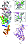
Figure 1
Figure 1
Crystal structure of TmTrpRS. (a) Schematic representation of the TmTrpRS crystallographic dimer. The α-helices are represented as cylinders in gray, β-strands as arrows in purple and the loops in pink in one monomer (top) of the dimer. The crystallographically related molecule below is color-coded from blue at the N-­terminus to red at the C-terminus. The bound 4Fe–4S cluster and l-tryptophan are represented as sticks. The β-strands (β1–β5) and α-helices (α1–α15) are labeled (310-helices are not labeled). The ATP-binding motifs (HIGH and KMSKS) are highlighted in red. The short hinge region (residues 182–186) connecting the Rossmann-fold and the TAB domains is indicated. (b) Close-up view of a 2F oF c OMIT map contoured at 1σ showing the iron–sulfur cluster bound to the cysteine motif [Cys236-x 22-Cys259-x 6-Cys266-x 2-Cys269] in the anticodon-binding region (top box) and the l-tryptophan found in the active site (bottom box). (c) Comparison of TmTrpRS to the ‘open’ and ‘closed’ conformations of BsTrpRS.
Figure 2
Figure 2
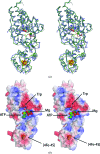
Comparison of TmTrpRS and DrTrpRS_II. (a) Structural superposition of TmTrpRS (gray), DrTrpRS_II + ATP and Mg (PDB code 1yid; green) and DrTrpRS_II + Trp (PDB code 1yi8; blue). The bound ATP is shown as a stick model. The [4Fe–4S] cluster (orange and yellow), Mg ion (green sphere) and Trp (gray, red and blue) are shown as spheres. The DrTrpRS_II structures superimpose with TmTrpRS over 319 Cα atoms with an r.m.s.d. of ∼1.7 Å (45% sequence identity). (b) Electrostatic surface potential of TmTrpRS calculated with the program APBS (Baker et al., 2001 ▶): positive potential is shown in blue (+3kTe−1) and negative in red (−3kTe−1). l-Tryptophan and the iron–sulfur cluster are shown as spheres. Note: the ATP-binding site is solvent-exposed, but the tryptophan and the iron–sulfur cluster are partially buried.
Figure 3
Figure 3
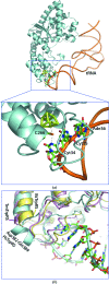
Model of the TmTrpRS–tRNA complex. (a) Overall view of the complex. TmTrpRS is shown as a gray-blue ribbon and tRNA as an orange trace. The anticodon bases, Cys266 and the [4Fe–4S] cluster are shown as stick models. Close-up view of the interaction of TrpRS–tRNA with the anticodon (CCA) region. Cyt34 of the tRNA molecule interacts with the iron of the cluster via Cys266. The model was based on the structure of the human TrpRS–tRNA complex (PDB code 1r6t). (b) Close-up view of the loop conformation of TmTrpRS (pink) near the [4Fe–4S] cluster (pink sticks) compared with the helix (Asp382–Asn389) of the human TrpRS–tRNA complex (gray–blue) and BsTrpRS (yellow).
Figure 4
Figure 4
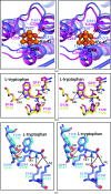
Iron–sulfur cluster and l-tryptophan recognition of TmTrpRS and comparison with other TrpRSs. (a) Comparison of the TmTrpRS (magenta) and human TrpRS (blue) TAB domains. The TmTrpRS iron–sulfur cluster is shown in orange ball-and-stick representation. (b) Comparison of the l-tryptophan recognition of TmTrpRS (magenta) and BsTrpRS (yellow). (c) Comparison of the l-tryptophan recognition of human TrpRS (blue) and ScTrpRS (slate blue).
Figure 5
Figure 5
Enzymatic activities of TmTrpRS. (a) Aminoacylation activity assayed at 310 and 333 K. Consistent with its thermophilic nature, TmTrpRS has a more robust tRNA-charging activity at 333 K compared with that at 310 K. Control reaction assays lacking enzyme or tRNA at 333 K are also shown. Points are the mean of two assays and error bars represent the standard error of the mean of measurements. (b) ATP–PPi-exchange activities assayed at 310 and 333 K. This experiment was only performed once. Plots were derived by fitting to an exponential rise to maximum function. Control reaction assays lacking enzyme or Trp at 333 K are also shown. Consistent with the observation of an endogenously bound Trp molecule in the active site of its crystal structure, TmTrpRS had some PPi-exchange activity even when no Trp was added to the reaction.
Figure 6
Figure 6
Structure-based sequence alignment of TrpRSs. The four Cys residues in the C-x 22-C-x 6-C-x 2-C motif of TmTrpRS that chelate the [4Fe–4S] cluster are colored by red letters. Strictly conserved residues are highlighted in red boxes and additional conserved residues are in yellow boxes. The secondary-structure elements of TmTrpRS are shown at the top, where α-­helices (α1–α15), β-strands (β1–β5) and 310-helices (η1–η4) are sequentially labeled and β-turns and γ-turns are designated with the Greek letter tau (T). Asp136, which is conserved among prokaryotes, is indicated with a blue diamond. Abbreviations: Thermotoga maritima TrpRS, TmTrpRS; Deinoccoccus radiodurans TrpRS_I, Dr_I; Bacillus stearothermophilus TrpRS, Bs; D. radiodurans TrpRS_II, Dr_II; Homo sapiens TrpRS, Hs. Note: the sequence of DrTrpRS_I is included in the alignment although there is no crystal structure available from the PDB.

References

    1. Altschul, S. F., Madden, T. L., Schäffer, A. A., Zhang, J., Zhang, Z., Miller, W. & Lipman, D. J. (1997). Nucleic Acids Res.25, 3389–3402. - PMC - PubMed
    1. Baker, N. A., Sept, D., Joseph, S., Holst, M. J. & McCammon, J. A. (2001). Proc. Natl Acad. Sci. USA, 98, 10037–10041. - PMC - PubMed
    1. Buddha, M. R. & Crane, B. R. (2005a). J. Biol. Chem.280, 31965–31973. - PubMed
    1. Buddha, M. R. & Crane, B. R. (2005b). Nature Struct. Mol. Biol.12, 274–275. - PubMed
    1. Chen, V. B., Arendall, W. B., Headd, J. J., Keedy, D. A., Immormino, R. M., Kapral, G. J., Murray, L. W., Richardson, J. S. & Richardson, D. C. (2010). Acta Cryst. D66, 12–21. - PMC - PubMed

Publication types

Associated data