. 2010 Oct 1;66(Pt 10):1354-64.
doi: 10.1107/S1744309110021214. Epub 2010 Jul 27.

Structure of the γ-D-glutamyl-L-diamino acid endopeptidase YkfC from Bacillus cereus in complex with L-Ala-γ-D-Glu: insights into substrate recognition by NlpC/P60 cysteine peptidases

Affiliations

Structure of the γ-D-glutamyl-L-diamino acid endopeptidase YkfC from Bacillus cereus in complex with L-Ala-γ-D-Glu: insights into substrate recognition by NlpC/P60 cysteine peptidases

Qingping Xu et al. Acta Crystallogr Sect F Struct Biol Cryst Commun. .

Abstract

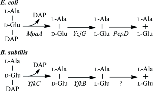
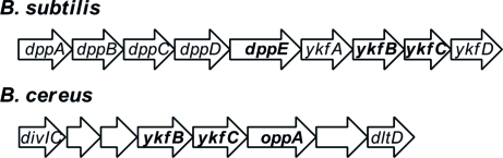
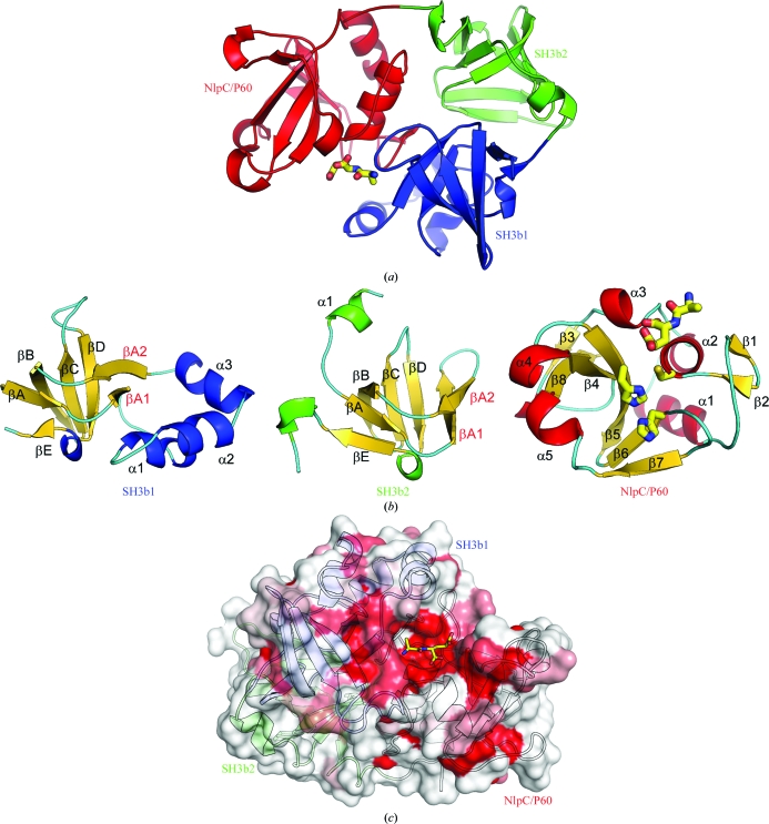
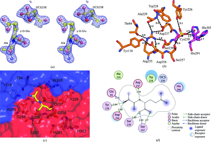
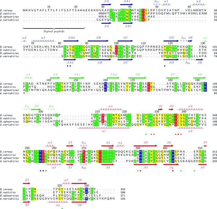
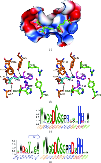
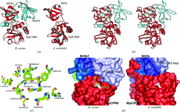
Dipeptidyl-peptidase VI from Bacillus sphaericus and YkfC from Bacillus subtilis have both previously been characterized as highly specific γ-D-glutamyl-L-diamino acid endopeptidases. The crystal structure of a YkfC ortholog from Bacillus cereus (BcYkfC) at 1.8 Å resolution revealed that it contains two N-terminal bacterial SH3 (SH3b) domains in addition to the C-terminal catalytic NlpC/P60 domain that is ubiquitous in the very large family of cell-wall-related cysteine peptidases. A bound reaction product (L-Ala-γ-D-Glu) enabled the identification of conserved sequence and structural signatures for recognition of L-Ala and γ-D-Glu and, therefore, provides a clear framework for understanding the substrate specificity observed in dipeptidyl-peptidase VI, YkfC and other NlpC/P60 domains in general. The first SH3b domain plays an important role in defining substrate specificity by contributing to the formation of the active site, such that only murein peptides with a free N-terminal alanine are allowed. A conserved tyrosine in the SH3b domain of the YkfC subfamily is correlated with the presence of a conserved acidic residue in the NlpC/P60 domain and both residues interact with the free amine group of the alanine. This structural feature allows the definition of a subfamily of NlpC/P60 enzymes with the same N-terminal substrate requirements, including a previously characterized cyanobacterial L-alanine-γ-D-glutamate endopeptidase that contains the two key components (an NlpC/P60 domain attached to an SH3b domain) for assembly of a YkfC-like active site.


Figures

Figure 1
Figure 1
Proposed metabolic pathways for murein peptides in E. coli and B. subtilis.
Figure 2
Figure 2
Genomic context of the ykfB and ykfC genes in B. subtilis and B. cereus.
Figure 3
Figure 3
Crystal structure of YkfC from B. cereus in complex with l-Ala-γ-d-Glu. (a) Ribbon representation of BcYkfC, highlighting its domain organization. SH3b1 is depicted in blue, SH3b2 in green and NlpC/P60 in red. The bound l-Ala-γ-d-Glu is shown as a stick model. (b) Ribbon representations of individual domains, showing the secondary-structure elements. (c) Molecular surface of YkfC colored by sequence conservation. The surface color gradient indicates the level of sequence conservation from the most conserved residues (deep red) to nonconserved residues (white).
Figure 4
Figure 4
Active site and recognition of l-Ala-γ-d-Glu by BcYkfC. (a) Stereoview of a 2F oF c OMIT map, where l-Ala-γ-d-Glu and OCS238 were omitted from phasing/refinement, contoured at 1.5σ. (b) The extensive hydrogen-bond network in the active site of BcYkfC. Hydrogen bonds and distances are shown as dashed lines. (c) l-Ala-γ-d-Glu (stick representation; yellow C atoms) is located in the active site at the interface of the SH3b1 domain (blue) and the NlpC/P60 domain (red). (d) The interaction between l-Ala-γ-d-Glu and the active site of YkfC. This figure was generated using the program MOE 2008.10 (Chemical Computing Group Inc.).
Figure 5
Figure 5
Sequence alignment of YkfC from B. subtilis and B. cereus, dipeptidyl-peptidase VI (DPP VI) from B. sphaericus and a γ-d-glutamyl-l-diamino acid endopeptidase from A. variabilis (AvPCP). The sequence numbering and secondary-structure elements of YkfC from B. cereus and AvPCP are indicated at the top and bottom, respectively. The alignment was generated by merging and manually editing the structure-based sequence alignment of BcYkfC and AvPCP with the sequence alignment of the top three sequences. The active-site residues are marked with colored dots at the bottom (blue, S2; orange, S1; red, catalytic triad; green, potential S′ sites).
Figure 6
Figure 6
Models of substrate recognition by BcYkfC. (a) l-Ala-γ-d-Glu-DAP was docked into the active site of BcYkfC. The protein surface is colored according to a gradient in electrostatic potential from negative (red) to positive (blue) (MOE 2008.10; Chemical Computing Group Inc.). (b) Stereoview of the specific interactions (four polar, one nonpolar) of γ-d-Glu (cyan) in the context of the tripeptide by five residues of YkfC (Tyr226, Trp228, Asp237, Ser239 and Ser257). The protein residues are colored according to subsite (S1, orange; catalytic triad, magenta; S1′, green). (c) Sequence conservation of the active sites in NlpC/P60 domains based on 2277 NlpC/P60 domains with an intact catalytic dyad (Cys238 and His291) and a conserved Tyr226 (blue, S2; orange, S1; red, catalytic triad; green, S′ sites). (d) Sequence conservation of the active sites in the YkfC subfamily of NlpC/P60 enzymes based on 282 sequences selected based on the presence of a conserved aspartate residue at position 256 of BcYkfC. The conservation of this residue is highly correlated with a conserved tyrosine in the βD region of the SH3b domain (Tyr118 of BcYkfC); both of these residues interact with the free amine of l-Ala of the substrate.
Figure 7
Figure 7
Structural comparisons between BcYkfC and the cyanobacterial NlpC/P60 endopeptidase AvPCP. (a) BcYkfC and AvPCP (PDB code 2hbw; Xu et al., 2009 ▶) are shown with the same orientation of their common SH3b1 and NlpC/P60 domains. The structurally equivalent residues in each are shown in red. (b) Stereoview of the Cα traces of the two proteins shown in (a) [the coloring is the same as in (a)]. (c) The S binding sites of the two proteins are nearly identical. The corresponding residues of AvPCP are labeled in parentheses. (d) Comparison of the active-site cavities and their environments. The catalytic cysteine is shown in white. A stick model of a docked murein tripeptide is shown in the active site of BcYkfC.

References

    1. Altschul, S. F., Madden, T. L., Schäffer, A. A., Zhang, J., Zhang, Z., Miller, W. & Lipman, D. J. (1997). Nucleic Acids Res.25, 3389–3402. - PMC - PubMed
    1. Anantharaman, V. & Aravind, L. (2003). Genome Biol.4, R11. - PMC - PubMed
    1. Aramini, J. M., Rossi, P., Huang, Y. J., Zhao, L., Jiang, M., Maglaqui, M., Xiao, R., Locke, J., Nair, R., Rost, B., Acton, T. B., Inouye, M. & Montelione, G. T. (2008). Biochemistry, 47, 9715–9717. - PubMed
    1. Asano, S. I., Nukumizu, Y., Bando, H., Iizuka, T. & Yamamoto, T. (1997). Appl. Environ. Microbiol.63, 1054–1057. - PMC - PubMed
    1. Bateman, A., Coin, L., Durbin, R., Finn, R. D., Hollich, V., Griffiths-Jones, S., Khanna, A., Marshall, M., Moxon, S., Sonnhammer, E. L., Studholme, D. J., Yeats, C. & Eddy, S. R. (2004). Nucleic Acids Res.32, D138–D141. - PMC - PubMed

Publication types

Substances

Associated data