Review
. 2010 Spring-Summer;7(2-3):e56-68.

An update on idiopathic intracranial hypertension

Affiliations
Review

An update on idiopathic intracranial hypertension

Matthew J Thurtell et al. Rev Neurol Dis. 2010 Spring-Summer.

Abstract

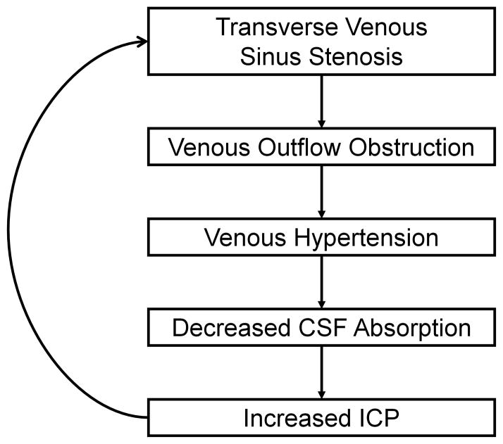
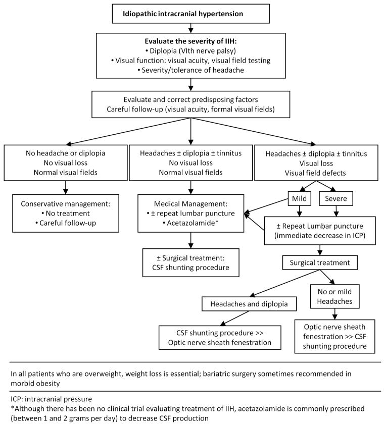
Idiopathic intracranial hypertension (IIH) is a condition of unknown etiology often encountered in neurologic practice. It produces nonlocalizing symptoms and signs of raised intracranial pressure and, when left untreated, can result in severe irreversible visual loss. It most commonly occurs in obese women of childbearing age, but it can also occur in children, men, nonobese adults, and older adults. Although it is frequently associated with obesity, it can be associated with other conditions, such as obstructive sleep apnea and transverse cerebral venous sinus stenoses. Recent identification of subgroups at high risk for irreversible visual loss, including black patients, men, and patients with fulminant forms of IIH, help guide the optimal management and follow-up. Ongoing studies of venous anatomy and physiology in IIH patients, as well as a recently begun randomized clinical treatment trial, should provide further insight into this common yet poorly understood syndrome.


Figures

Figure 1
Figure 1. Papilledema and subtle visual field changes in an obese young woman with IIH
A: At presentation, the patient had severe headache, bilateral papilledema (elevated optic nerves, peripapillary hemorrhages, and dilated retinal veins), and enlargement of the physiologic blind spots on 24-2 Humphrey visual fields. Following a diagnostic lumbar puncture, her headache improved for several days. She had refused to take acetazolamide, but attempted to lose weight. B: The patient’s headaches and papilledema worsened, despite one lumbar puncture and attempts at weight loss, but her visual fields remained stable. Since she reported dramatic, but transient, improvement in symptoms following serial lumbar punctures, she was referred for a CSF shunting procedure. C: Three months following lumbo-peritoneal shunting, her symptoms and signs had resolved. The optic nerves remained mildly elevated, with mild gliosis and pallor, but her visual fields were full.
Figure 2
Figure 2. Fulminant onset IIH, with severe headaches, bilateral papilledema, and visual loss
A: At the time of presentation, the patient had severe papilledema, with exudates, decreased visual acuity in both eyes, and severely constricted Goldmann visual fields in both eyes. Her headaches dramatically improved for a few hours after the diagnostic lumbar puncture. She worsened over the next 24 hours and received a lumbar drain, followed by an urgent CSF shunting procedure. B: Despite shunting, the patient developed secondary optic atrophy in both eyes and had persistent severe visual field loss.
Figure 3
Figure 3. Imaging findings in IIH
A: Empty sella (*) on sagittal T1-weighted imaging. B: Dilated optic nerve sheaths (arrowheads) on axial T2-weighted imaging. C: Posterior globe flattening (arrowheads) and enhancement of protruding optic nerve heads (arrows) on axial T1-weighted imaging with contrast.
Figure 4
Figure 4. Transverse cerebral venous sinus stenosis in an IIH patient, as seen on contrast-enhanced MR venography. There is a long narrow stenosis of the right transverse venous sinus (arrowheads) and a hypoplastic left transverse venous sinus
A: Sagittal plane. B: Coronal plane. C: Axial plane.
Figure 5
Figure 5
Schematic summary of the presumed role of transverse venous sinus stenosis in IIH.
Figure 6
Figure 6
Algorithm summarizing our suggested approach to IIH management, including conservative, medical, and surgical treatments (reprinted with permission from Biousse and Newman, 2009).

References

    1. Friedman DI, Jacobson DM. Diagnostic criteria for idiopathic intracranial hypertension. Neurology. 2002;59:1492–1495. - PubMed
    1. Biousse V, Ameri A, Bousser MG. Isolated intracranial hypertension as the only sign of cerebral venous thrombosis. Neurology. 1999;53:1537–1542. - PubMed
    1. Digre KB, Corbett JJ. Idiopathic intracranial hypertension (pseudotumor cerebri): a reappraisal. Neurologist. 2001;7:2–67.
    1. Friedman DI, Jacobson DM. Idiopathic intracranial hypertension. J Neuroophthalmol. 2004;24:138–145. - PubMed
    1. Digre KB. Three current controversies in idiopathic intracranial hypertension. Neuroophthalmology. 2009;33:93–99.

Publication types

LinkOut - more resources