

Electroconvulsive therapy alters dopamine signaling in the striatum of non-human primates
- PMID: 20944554
- PMCID: PMC3055667
- DOI: 10.1038/npp.2010.182
Electroconvulsive therapy alters dopamine signaling in the striatum of non-human primates
Abstract
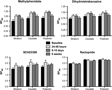
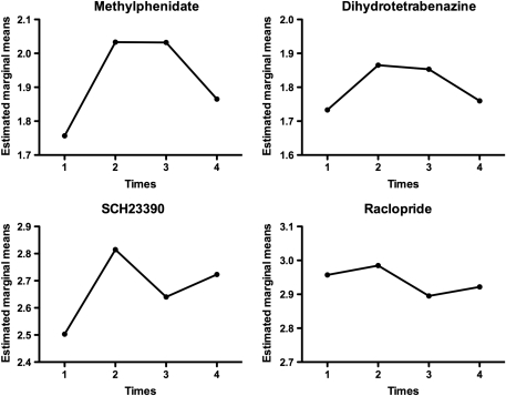
Electroconvulsive therapy (ECT) is one of the most effective therapies for depression and has beneficial motor effects in parkinsonian patients. However, little is known about the mechanisms of therapeutic action of ECT for either condition. The aim of this work was to explore the impact of ECT on dopaminergic function in the striatum of non-human primates. Rhesus monkeys underwent a course of six ECT treatments under a human clinical protocol. Longitudinal effects on the dopaminergic nigrostriatal system were studied over 6 weeks using the in vivo capabilities of positron emission tomography (PET). PET scans were performed prior to the onset of ECT treatments and at 24-48 h, 8-10 days, and 6 weeks after the final ECT treatment. Early increases in dopamine transporter and vesicular monoamine transporter 2 binding returned to baseline levels by 6 weeks post-ECT. Transient increases in D1 receptor binding were also observed, whereas the binding potential to D2 receptors was unaltered. The increase in dopaminergic neurotransmission suggested by our results may account in part for the therapeutic effect of ECT in mood disorders and Parkinson's disease.
Figures
Similar articles
-
Antiparkinsonian mechanism of electroconvulsive therapy in MPTP-lesioned non-human primates.Neurodegener Dis. 2012;9(3):128-38. doi: 10.1159/000334497. Epub 2012 Feb 9. Neurodegener Dis. 2012. PMID: 22327563
-
Electroconvulsive therapy decreases striatal dopamine transporter binding in patients with depression: A positron emission tomography study with [18F]FE-PE2I.Psychiatry Res Neuroimaging. 2020 Jul 30;301:111086. doi: 10.1016/j.pscychresns.2020.111086. Epub 2020 May 4. Psychiatry Res Neuroimaging. 2020. PMID: 32464340
-
Elevated dopamine D1 receptor availability in striatum of Göttingen minipigs after electroconvulsive therapy.J Cereb Blood Flow Metab. 2018 May;38(5):881-887. doi: 10.1177/0271678X17705260. Epub 2017 May 16. J Cereb Blood Flow Metab. 2018. PMID: 28509598 Free PMC article.
-
Electroconvulsive therapy (ECT) in Parkinson's disease: ECS and dopamine enhancement.J ECT. 2014 Jun;30(2):122-4. doi: 10.1097/YCT.0000000000000142. J ECT. 2014. PMID: 24810775 Review.
-
Postsynaptic nigrostriatal dopamine receptors and their role in movement regulation.J Neural Transm (Vienna). 2010 Dec;117(12):1359-69. doi: 10.1007/s00702-010-0454-z. Epub 2010 Nov 16. J Neural Transm (Vienna). 2010. PMID: 21076988 Free PMC article. Review.
Cited by
-
Electroconvulsive Treatment: Hypotheses about Mechanisms of Action.Front Psychiatry. 2013 Aug 27;4:94. doi: 10.3389/fpsyt.2013.00094. eCollection 2013. Front Psychiatry. 2013. PMID: 23986724 Free PMC article.
-
Moderate-level prenatal alcohol exposure induces sex differences in dopamine d1 receptor binding in adult rhesus monkeys.Alcohol Clin Exp Res. 2014 Dec;38(12):2934-43. doi: 10.1111/acer.12575. Alcohol Clin Exp Res. 2014. PMID: 25581649 Free PMC article.
-
Antidepressant treatment effects on dopamine transporter availability in patients with major depression: a prospective 123I-FP-CIT SPECT imaging genetic study.J Neural Transm (Vienna). 2018 Jun;125(6):995-1005. doi: 10.1007/s00702-018-1863-7. Epub 2018 Feb 23. J Neural Transm (Vienna). 2018. PMID: 29476250
-
Beyond neural cubism: promoting a multidimensional view of brain disorders by enhancing the integration of neurology and psychiatry in education.Acad Med. 2015 May;90(5):581-6. doi: 10.1097/ACM.0000000000000530. Acad Med. 2015. PMID: 25340364 Free PMC article. Review.
-
Molecular Biomarkers of Electroconvulsive Therapy Effects and Clinical Response: Understanding the Present to Shape the Future.Brain Sci. 2021 Aug 25;11(9):1120. doi: 10.3390/brainsci11091120. Brain Sci. 2021. PMID: 34573142 Free PMC article. Review.
References
-
- Andersen K, Balldin J, Gottfries CG, Granerus AK, Modigh K, Svennerholm L, et al. A double-blind evaluation of electroconvulsive therapy in Parkinson's disease with ‘on-off' phenomena. Acta Neurol Scand. 1987;76:191–199. - PubMed
-
- Antonini A, Schwarz J, Oertel WH, Beer HF, Madeja UD, Leenders KL. [11C]-Raclopride and positron emission tomography in previously untreated patients with Parkinson's disease: influence of L-dopa and lisuride therapy on striatal dopamine D2-receptors. Neurology. 1994;44:1325–1329. - PubMed
-
- Barkai AI, Durkin M, Nelson HD. Localized alterations of dopamine receptor binding in rat brain by repeated electroconvulsive shock: an autoradiographic study. Brain Res. 1990;529:208–213. - PubMed
-
- Bergstrom DA, Kellar KJ. Effect of electroconvulsive shock on monoaminergic receptor binding sites in rat brain. Nature. 1979;278:464–466. - PubMed
-
- Brown R, Jahanshahi M. Depression in Parkinson's disease: a psychosocial viewpoint. Adv Neurol. 1995;65:61–84. - PubMed
Publication types
MeSH terms
Substances
Grants and funding
LinkOut - more resources
Full Text Sources
Other Literature Sources
