Review
. 2010 Nov;11(11):760-72.
doi: 10.1038/nrn2915. Epub 2010 Oct 14.

Goal-directed and habitual control in the basal ganglia: implications for Parkinson's disease

Affiliations
Review

Goal-directed and habitual control in the basal ganglia: implications for Parkinson's disease

Peter Redgrave et al. Nat Rev Neurosci. 2010 Nov.

Abstract

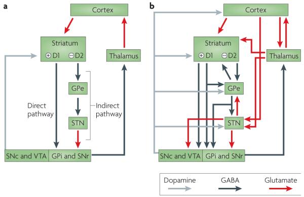
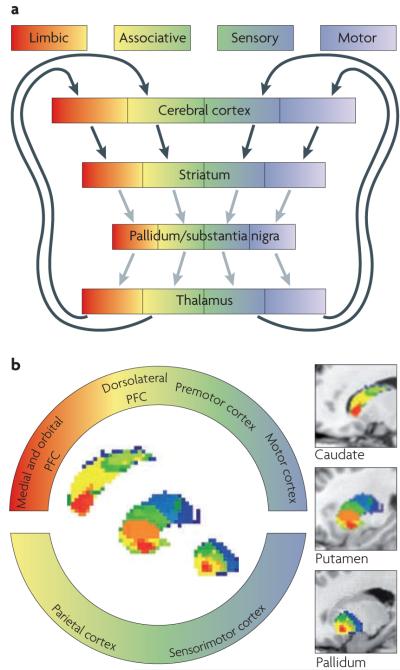
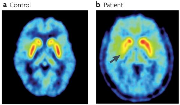
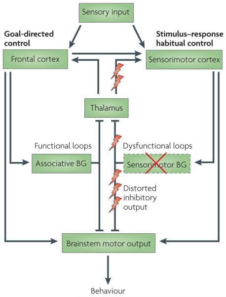
Progressive loss of the ascending dopaminergic projection in the basal ganglia is a fundamental pathological feature of Parkinson's disease. Studies in animals and humans have identified spatially segregated functional territories in the basal ganglia for the control of goal-directed and habitual actions. In patients with Parkinson's disease the loss of dopamine is predominantly in the posterior putamen, a region of the basal ganglia associated with the control of habitual behaviour. These patients may therefore be forced into a progressive reliance on the goal-directed mode of action control that is mediated by comparatively preserved processing in the rostromedial striatum. Thus, many of their behavioural difficulties may reflect a loss of normal automatic control owing to distorting output signals from habitual control circuits, which impede the expression of goal-directed action.


Figures

Figure 1
Figure 1. Organization of intrinsic connections within the basal ganglia
a ∣ Model based on the influential proposal by Albin and colleagues, according to which the output of the basal ganglia is determined by the balance between the direct pathway — which involves direct striatonigral inhibitory connections that promote behaviour — and the indirect pathway — which involves relays in the external globus pallidus (GPe) and subthalamic nucleus (STN), and suppresses behaviour. The balance between these two projections was thought to be regulated by afferent dopaminergic signals from the substantia nigra pars compacta (SNc) and the ventral tegmental area (VTA) acting on differentially distributed D1 and D2 dopamine receptors. b ∣ Recent anatomical investigations have revealed a rather more complex organization in which the transformations that are applied to the inputs to generate outputs are less easy to predict. GPe, globus pallidus pars externa; GPi, globus pallidus pars interna; SNr, substantia nigra pars reticulata. Figure is modified from REF. © P. Redgrave (2007).
Figure 2
Figure 2. Corticobasal ganglia–cortical loops in animals and humans
a ∣ The connections between the cerebral cortex and the basal ganglia can be viewed as a series of parallel-projecting, largely segregated loops or channels conveying limbic (shown in red), associative (shown in yellow–green) and sensorimotor (shown in blue–white) information. Functional territories represented at the level of cerebral cortex are maintained throughout the basal ganglia nuclei and thalamic relays. Note, however, that for each loop the relay points in the cortex, basal ganglia and thalamus, offer opportunities for activity inside the loop to be modified or modulated by signals from outside the loop. Black arrows indicate glutamatergic projections, grey arrows indicate GABA (γ-aminobutyric acid)-ergic projections. b ∣ The spatially segregated ‘rostral caudal gradient’ of human frontal cortical connectivity in caudate, putamen and pallidum. The colour-coded ring denotes limbic (shown in red), associative (shown in yellow–green) and sensorimotor regions of the cerebral cortex in the sagittal plane (colour coding after Haber173). Using probabilistic tractography on magnetic resonance-diffusion weighted imaging data, Draganski et al. identified the regions of the striatum that receive the strongest input from the identified cortical regions (sagittal oriented images contained within the ring). For better visualization, segmented basal ganglia nuclei are superimposed on T1-weighted structural images in the far right of the diagram (the rostral part is shown on the left, the caudal part is shown on the right). PFC, prefrontal cortex. Part a is modified, with permission, from REF. © (1995) MIT Press. Part b is reproduced, with permission, from REF. © (2008) Society for Neuroscience.
Figure 3
Figure 3. Striatal determinants of goal-directed and habitual action in rodents and humans
a ∣ Photomicrographs of NMDA (N-methyl-d-aspartate)-induced cell-body lesions of the rat dorsomedial and dorsolateral striatum (shown in the right hemisphere) with approximate regions of lesion-induced damage illustrated with red and purple circles, respectively (shown in the left hemisphere). Lesions of the dorsomedial striatum abolished acquisition and retention of goal-directed learning, whereas lesions of dorsolateral striatum abolished the acquisition of habit learning. b ∣ The regions of the human anterior dorsomedial striatum that exhibits sensitivity to instrumental contingency. c ∣ The region of the human posterior lateral striatum (posterior putamen) that exhibits a response profile that is consistent with the development of habits in humans. Part a is reproduced, with permission, from REF. © (2010) Macmillan Publishers Ltd. All rights reserved. Part b is reproduced, with permission, from REF. © (2008) Society for Neuroscience. Part c is reproduced, with permission, from REF. © (2009) Blackwell Publishing Ltd.
Figure 4
Figure 4. Striatal dopamine innervation assessed by 18fluorodopa positron emission tomography (PET)
a ∣ PET scan from a control subject showing high uptake of 18fluorodopa in the striatum (highest uptake values are shown in white). b ∣ An example of a PET scan from a patient with Parkinson’s disease with motor signs that are mainly confined to the right limbs. 18Fluorodopa uptake is markedly reduced in the left posterior putamen (the uptake in the area indicated by the arrow is 70% below normal) and reduced to a minor extent in the anterior putamen and caudate of the left hemisphere.
Figure 5
Figure 5. Functional and dysfunctional loops through the basal ganglia in the parkinsonian state
Both goal-directed and habitual control systems receive sensory inputs. Goal-directed control is dependent on the associative networks through the basal ganglia, whereas stimulus–response habitual control relies on the sensorimotor territories. Both networks independently have the capacity to direct behavioural output via ‘final common motor pathways’ (REFS 84,85,87). There may be many sites at which output from the two control systems might converge, and two have been illustrated: one at the level of cortical motor output, the other in the brainstem. In Parkinson’s disease, differential loss of dopamine innervation from sensorimotor regions in the basal ganglia (shown by the red cross) causes dysfunctional output signals from these territories and their associated networks (shown by lightning symbols). Presumably, this loss of function causes an increased reliance on goal-directed control. In addition, the distorting inhibitory output from sensorimotor territories is likely to impede the execution of goal-directed behavioural control that is otherwise comparatively preserved. BG, basal ganglia.

References

    1. Ferrier D. The Functions of the Brain. Putnam’s Sons; New York: 1876.
    1. Penney JB, Jr, Young AB. Striatal inhomogeneities and basal ganglia function. Mov. Disord. 1986;1:3–15. - PubMed
    1. Albin RL, Young AB, Penney JB. The functional anatomy of basal ganglia disorders. Trends Neurosci. 1989;12:366–375. - PubMed
    1. Chevalier G, Deniau JM. Disinhibition as a basic process in the expression of striatal functions. Trends Neurosci. 1990;13:277–281. This review introduced a conceptual development in suggesting that a pause in neuronal firing in basal ganglia output nuclei disinhibits efferent targets and is the major physiological mechanism by which the basal ganglia exert their effects on behaviour.

    1. Gerfen CR, Wilson CJ. In: Handbook of Chemical Neuroanatomy. Swanson LW, Bjorklund A, Hokfelt T, editors. Vol 12. Elsevier; Amsterdam: 1996. pp. 371–468.

Publication types