. 2010 Oct 14;467(7317):805-10.
doi: 10.1038/nature09423.

The Ndc80 kinetochore complex forms oligomeric arrays along microtubules

Affiliations

The Ndc80 kinetochore complex forms oligomeric arrays along microtubules

Gregory M Alushin et al. Nature. .

Abstract

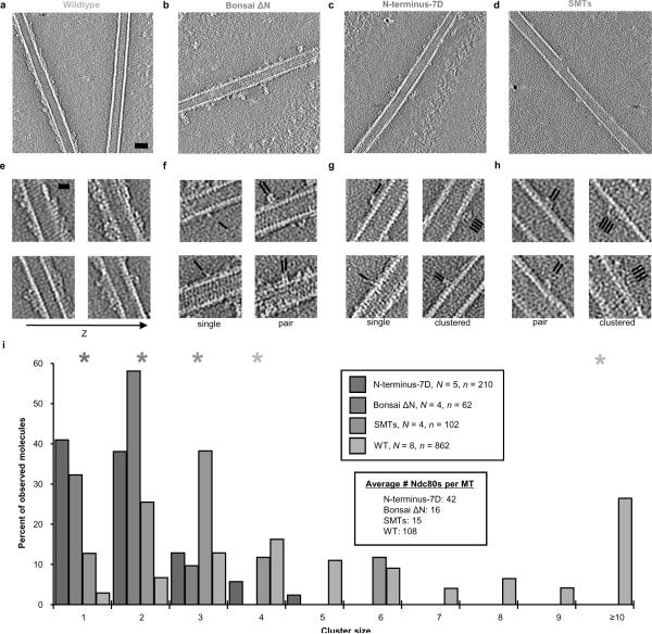
The Ndc80 complex is a key site of regulated kinetochore-microtubule attachment (a process required for cell division), but the molecular mechanism underlying its function remains unknown. Here we present a subnanometre-resolution cryo-electron microscopy reconstruction of the human Ndc80 complex bound to microtubules, sufficient for precise docking of crystal structures of the component proteins. We find that the Ndc80 complex binds the microtubule with a tubulin monomer repeat, recognizing α- and β-tubulin at both intra- and inter-tubulin dimer interfaces in a manner that is sensitive to tubulin conformation. Furthermore, Ndc80 complexes self-associate along protofilaments through interactions mediated by the amino-terminal tail of the NDC80 protein, which is the site of phospho-regulation by Aurora B kinase. The complex's mode of interaction with the microtubule and its oligomerization suggest a mechanism by which Aurora B could regulate the stability of load-bearing kinetochore-microtubule attachments.


Figures

Figure 1
Figure 1. Structure of the Ndc80-microtubule interface
a, End on (from the plus-end) and side views of the microtubule-Ndc80 complex cryo-EM reconstruction (tubulin, green; Ndc80-Nuf2 head, blue; disordered Spc24–25 head, red). b, Orthogonal views of docked crystal structures (Ndc80, blue; gold-Nuf2 gold; green-tubulin). In ball and stick: ordered residues adjacent to the N-terminus of Bonsai ΔN (magenta), ordered residues in tubulin preceding the E-hooks (red). The region of the map occupied by Nuf2 (right panel) is at higher radius and thus is of lower resolution. c, Orthogonal views of the positive difference density (magenta) between the cryo-EM reconstruction and the docked crystal structures, contoured at 2.5 σ. The dotted lines indicate a proposed path for the Ndc80 N-terminus.
Figure 2
Figure 2. The Ndc80 toe-print is a tubulin conformation sensor
a, Superposition of intra-dimer and inter-dimer interfaces, viewed from the outside of the microtubule (α-tubulin, green; β-tubulin, blue; ordered residues adjacent to the E-hooks, red; conserved toe-print segments, purple and orange). Consensus sequences are indicated, with deviations between the monomers in parenthesis. b, SDS-PAGE of co-sedimentation assays with straight (taxol) and curved (vinblastine) tubulin polymers with the indicated Ndc80 bonsai constructs. S: supernatant, P: pellet. [Tubulin monomer] = 6 μM, [Ndc80 bonsai] = 0.5 μM. c, Quantification of b. Error bars represent ± s.d., n = 3. d, SDS-PAGE of sedimentation assays after cold-induced depolymerization of dynamic microtubules in the presence and absence of Ndc80 bonsai. e, Negative-stain EM of Ndc80-induced cold-stable microtubules and straight tubulin sheets. Scale bar, 50 nm.
Figure 3
Figure 3. Cluster formation requires the N-terminus of the Ndc80 protein
a–d, Central slices of tomograms of microtubule-bound Ndc80 constructs under subsaturating conditions. Ndc80:tubulin monomer ration was 1:2 for wild-type and SMTs and 2:1 for bonsai N-terminus mutants. SMTs: subtilisin-cleaved microtubules. Scale bar, 25 nm (black dots = gold fiducials). e, Serial slices of the wild-type reconstruction 4.5 nm apart show cooperative binding and cluster formation only along (not between) protofilaments. Scale bar, 10 nm. f–h, Selected views of clusters in Bonsai ΔN, N-terminus-7D, and SMTs reconstructions, respectively. Black lines indicate position and orientation of Ndc80 molecules. i, Quantification of cluster size populations. N = number of reconstructed MT segments, n = total number of Ndc80 complexes observed. Asterisks indicate the most probable cluster size for each of the populations (wild-type has two peaks). See Supplementary Table 1 for pair-wise statistical comparison of these distributions.
Figure 4
Figure 4. Proposed models of attachment maturation and biased diffusion
a, A cartoon illustrating the phospho-regulated formation of Ndc80 clusters in vivo concomitant with stable kinetochore-mirotubule attachment (colors are as Fig. 1b). In this schematic the assembly of the cluster biased diffusion machinery is regulated by the spatial localization of Aurora B and a counterbalancing phosphatase rather than directly by tension. b, A cartoon diagramming the proposed biased diffusion process for coupling chromosome movement to microtubule depolymerization via the Ndc80 complex. Colors are as in Fig. 1b, except the Ndc80-Nuf2 head is shown in gold and unidentified filaments are shown in grey.

References

    1. Cheeseman IM, Chappie JS, Wilson-Kubalek EM, Desai A. The conserved KMN network constitutes the core microtubule-binding site of the kinetochore. Cell. 2006;127:983–997. - PubMed
    1. Wei RR, Al-Bassam J, Harrison SC. The Ndc80/HEC1 complex is a contact point for kinetochore-microtubule attachment. Nat Struct Mol Biol. 2007;14:54–59. - PubMed
    1. DeLuca JG, et al. Kinetochore microtubule dynamics and attachment stability are regulated by Hec1. Cell. 2006;127:969–982. - PubMed
    1. Martin-Lluesma S, Stucke VM, Nigg EA. Role of Hec1 in spindle checkpoint signaling and kinetochore recruitment of Mad1/Mad2. Science. 2002;297:2267–2270. - PubMed
    1. DeLuca JG, et al. Nuf2 and Hec1 are required for retention of the checkpoint proteins Mad1 and Mad2 to kinetochores. Curr Biol. 2003;13:2103–2109. - PubMed

Publication types

Associated data