. 2010 Dec;19(12):2389-401.
doi: 10.1002/pro.519.

Structural and thermodynamic analysis of the GFP:GFP-nanobody complex

Affiliations

Structural and thermodynamic analysis of the GFP:GFP-nanobody complex

Marta H Kubala et al. Protein Sci. 2010 Dec.

Abstract

The green fluorescent protein (GFP)-nanobody is a single-chain VHH antibody domain developed with specific binding activity against GFP and is emerging as a powerful tool for isolation and cellular engineering of fluorescent protein fusions in many different fields of biological research. Using X-ray crystallography and isothermal titration calorimetry, we determine the molecular details of GFP:GFP-nanobody complex formation and explain the basis of high affinity and at the same time high specificity of protein binding. Although the GFP-nanobody can also bind YFP, it cannot bind the closely related CFP or other fluorescent proteins from the mFruit series. CFP differs from GFP only within the central chromophore and at one surface amino acid position, which lies in the binding interface. Using this information, we have engineered a CFP variant (I146N) that is also able to bind the GFP-nanobody with high affinity, thus extending the toolbox of genetically encoded fluorescent probes that can be isolated using the GFP-nanobody.


Figures

Figure 1
Figure 1
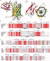
Structure of the GFP:GFP-nanobody heterodimeric complex determined by X-ray crystallography. (A) Ribbon diagram showing perpendicular views of the GFP:GFP-nanobody complex. GFP is shown in green to gold and GFP-nanobody is shown colored red to pink (N- to C-terminal). The GFP chromophore is shown in stick representation. (B) Sequence alignment of the GFP-nanobody with other camelid nanobodies of known structure. The CDR regions are boxed, and the characteristic disulfide bridge indicated. Side-chains that directly contact GFP are indicated by green triangles. (C) Sequence alignment of GFP with other fluorescent proteins. The three residues that are cyclized to form the chromophore are boxed in green. Side-chains that make direct contact with the GFP-nanobody are indicated by red triangles.
Figure 2
Figure 2
Details of the GFP:GFP-nanobody interface. (A) View of the binding interface. Residues participating in the interface are shown as sticks. Proteins are colored as in Figure 1. (B) Close up showing details of the environment surrounding GFP Asn146. Asn146 is the only side-chain in the interface that varies between GFP and CFP (Ile146 in CFP) [Fig. 1(D)] and, therefore, determines the nanobody specificity. Hydrogen bonds are indicated with dashed red lines.
Figure 3
Figure 3
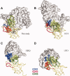
Comparison of the GFP:GFP-nanobody complex with other representative camelid nanobody protein complexes. The representative sequences of each nanobody are given in Figure 1(B). In each structure the nanobody is shown as a yellow worm with CDR1 colored blue, CDR2 colored red, and CDR3 colored green. The bound proteins are shown as surface representations with atoms that contact each CDR loop colored, respectively. (A) The GFP:GFP-nanobody complex. (B) The EpsI:EpsJ:nanobody complex (PDB ID 3CFI10). (C) The RNaseA:nanobody complex (PDB ID 1BXQ14). (D) The lysozyme:nanobody complex (PDB ID 1JTO17).
Figure 4
Figure 4
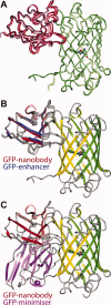
Comparison of the GFP:GFP-nanobody complex with GFP-enhancer and GFP-minimizer structures. (A) In the GFP:GFP-nanobody crystals four copies of the complex are present in the asymmetric unit. An overlay is shown of the four copies as Cα traces, with GFP in different shades of green and GFP-nanobody in shades of red. There is no significant difference between the four subunits. (B) Comparison of the GFP:GFP-nanobody structure (this study) colored as in Figure 1(A) with the GFP:GFP-enhancer structure (PDB ID 3K1K4). Only GFP from this study is shown for clarity, whereas the GFP-enhancer is shown in blue. (C) Comparison of the GFP:GFP-nanobody structure (this study) colored as in Figure 1(A) with the GFP:GFP-minimizer structure (PDB ID 3G9A4). Only GFP from this study is shown for clarity, while the GFP-minimizer is shown in purple.
Figure 5
Figure 5
Interaction analysis of the GFP:GFP-nanobody complex. (A) Binding of GFP to biotinylated GFP-nanobody measured using an Octet RED interferometer. Depicted sensorgrams represent complex formation (first 300 s) at 500, 250, 125, 62.5, 31.25, and 0 nM of GFP and subsequent dissociation of the complex in binding buffer without GFP (300–1200 s). (B) ITC binding thermogram of GFP binding to GFP-nanobody. GFP-nanobody (47 μM) was titrated into GFP (5.2 μM) at 37°C. The top panel shows the raw data, whereas the bottom panel shows the integrated normalized data for binding. (C) Graph of ΔH vs. temperature for the interaction of GFP with the GFP-nanobody. (D) Cut-away surface representations show the binding interfaces of GFP and GFP-nanobody. GFP is colored with red to highlight contact with GFP-nanobody, whereas GFP-nanobody is conversely colored green to indicate contact with GFP.
Figure 6
Figure 6
Engineering of CFP for GFP-nanobody binding. (A) Normalized fluorescence emission spectra for GFP, CFP, and mutant proteins after excitation at 430 nm. (B) Fluorescence intensity of GFP, CFP, and mutants measured using 430Ex/485Em and 485Ex/528Em filter sets, respectively. (C) Affinity precipitation of in vitro translated GFP, CFP, and their mutants were performed using sepharose-coupled GFP-nanobody, and the eluates were analyzed by SDS-PAGE followed by anti-GFP/CFP Western blotting (D) Quantification of gels shown in Figure 6(C) by IR densitometry (Odyssey Imaging System).

References

    1. Tsien RY. Constructing and exploiting the fluorescent protein paintbox (Nobel Lecture) Angew Chem Int Ed Engl. 2009;48:5612–5626. - PubMed
    1. Skerra A. Alternative non-antibody scaffolds for molecular recognition. Curr Opin Biotechnol. 2007;18:295–304. - PubMed
    1. Muyldermans S, Baral TN, Retamozzo VC, De Baetselier P, De Genst E, Kinne J, Leonhardt H, Magez S, Nguyen VK, Revets H, Rothbauer U, Stijlemans B, Tillib S, Wernery U, Wyns L, Hassanzadeh-Ghassabeh G, Saerens D. Camelid immunoglobulins and nanobody technology. Vet Immunol Immunopathol. 2009;128:178–183. - PubMed
    1. Kirchhofer A, Helma J, Schmidthals K, Frauer C, Cui S, Karcher A, Pellis M, Muyldermans S, Casas-Delucchi CS, Cardoso MC, Leonhardt H, Hopfner KP, Rothbauer U. Modulation of protein properties in living cells using nanobodies. Nat Struct Mol Biol. 2010;17:133–138. - PubMed
    1. Rothbauer U, Zolghadr K, Muyldermans S, Schepers A, Cardoso MC, Leonhardt H. A versatile nanotrap for biochemical and functional studies with fluorescent fusion proteins. Mol Cell Proteomics. 2008;7:282–289. - PubMed

Publication types

MeSH terms

Associated data