Review
. 2011 Jul;202(3):387-407.
doi: 10.1111/j.1748-1716.2010.02195.x. Epub 2010 Nov 16.

Transcellular and paracellular pathways of transepithelial fluid secretion in Malpighian (renal) tubules of the yellow fever mosquito Aedes aegypti

Affiliations
Review

Transcellular and paracellular pathways of transepithelial fluid secretion in Malpighian (renal) tubules of the yellow fever mosquito Aedes aegypti

K W Beyenbach et al. Acta Physiol (Oxf). 2011 Jul.

Abstract

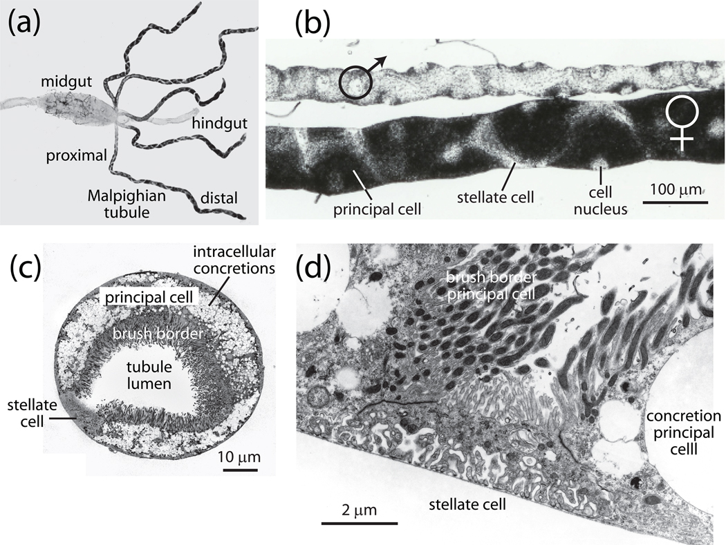
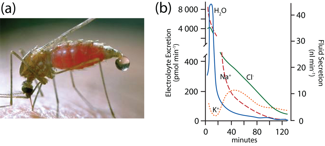
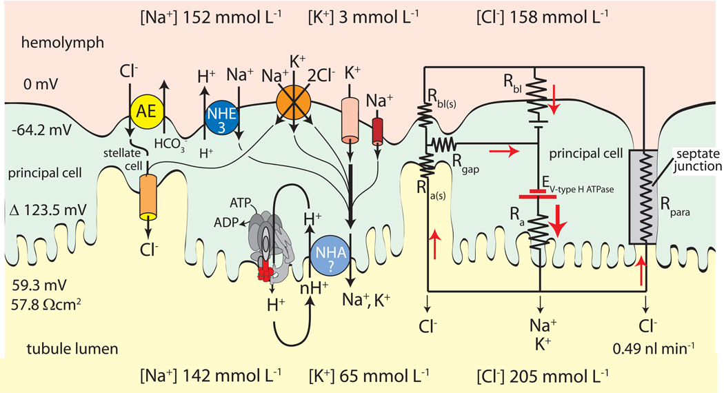
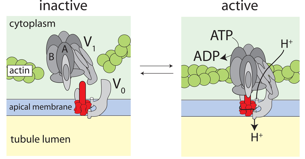
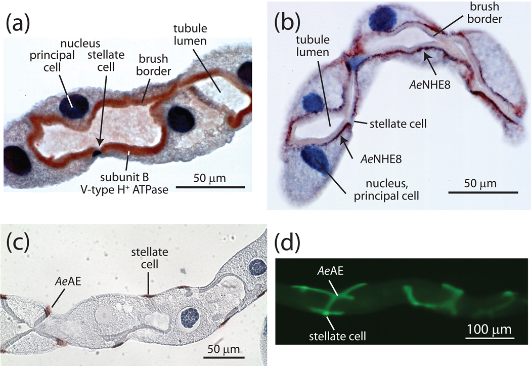
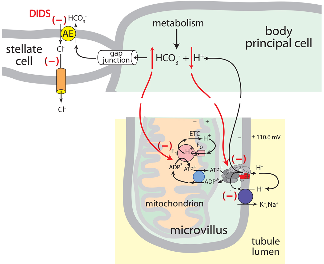
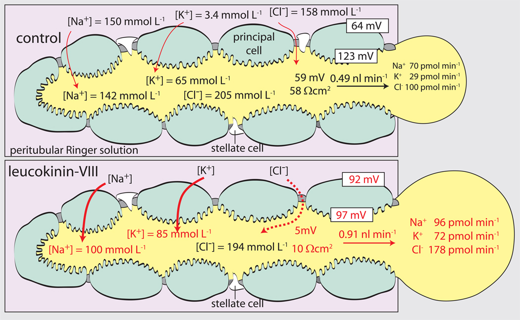
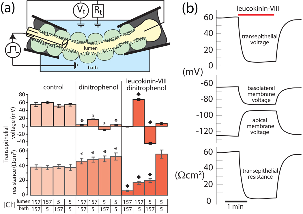
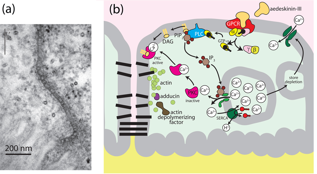
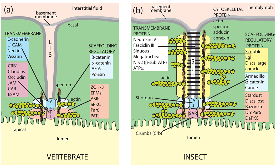
Isolated Malpighian tubules of the yellow fever mosquito secrete NaCl and KCl from the peritubular bath to the tubule lumen via active transport of Na(+) and K(+) by principal cells. Lumen-positive transepithelial voltages are the result. The counter-ion Cl(-) follows passively by electrodiffusion through the paracellular pathway. Water follows by osmosis, but specific routes for water across the epithelium are unknown. Remarkably, the transepithelial secretion of NaCl, KCl and water is driven by a H(+) V-ATPase located in the apical brush border membrane of principal cells and not the canonical Na(+), K(+) -ATPase. A hypothetical cation/H(+) exchanger moves Na(+) and K(+) from the cytoplasm to the tubule lumen. Also remarkable is the dynamic regulation of the paracellular permeability with switch-like speed which mediates in part the post-blood-meal diuresis in mosquitoes. For example, the blood meal the female mosquito takes to nourish her eggs triggers the release of kinin diuretic peptides that (i) increases the Cl(-) conductance of the paracellular pathway and (ii) assembles V(1) and V(0) complexes to activate the H(+) V-ATPase and cation/H(+) exchange close by. Thus, transcellular and paracellular pathways are both stimulated to quickly rid the mosquito of the unwanted salts and water of the blood meal. Stellate cells of the tubule appear to serve a metabolic support role, exporting the HCO(3)(-) generated during stimulated transport activity. Septate junctions define the properties of the paracellular pathway in Malpighian tubules, but the proteins responsible for the permselectivity and barrier functions of the septate junction are unknown.


Figures

Fig. 1
Fig. 1
Malpighian tubules of the yellow fever mosquito Aedes aegypti. A, five Malpighian tubules empty their secretions into the gut at the junction of the midgut and hindgut of an adult female mosquito (modified from (Yu and Beyenbach, 2004). B, sexual dimorphism of adult Malpighian tubules in the yellow fever mosquito. Principal cells are large and opaque on account of the density of intracellular concretions, and stellate cells are small and transparent (modified from (Plawner et al., 1991). C, transverse section through a Malpighian tubule of a female yellow fever mosquito. Note the tall, mitochondrion-rich brush border and the intracellular concretions in principal cells, and the small size of the stellate cell (modified from(Beyenbach and Piermarini, 2009). D, stellate cell with extensive infoldings of the basal membrane, a sparse cytoplasm and a short brush border. Note the brush border of neighboring principal cells where each microvillus is home to a mitochondrion (modified from (Beyenbach and Piermarini, 2009).
Fig. 2
Fig. 2
The blood meal diuresis in two mosquitoes. A, the diuresis in the mosquito Anopheles freeborni (courtesy of Jack Kelly Clark, University of California). Note the onset of the diuresis before the blood meal has been finished. B, in vivo time course of the blood meal diuresis in the yellow fever mosquito Aedes aegypti. The droplets ejected from the rectum were analyzed for the composition of Na+, K+ and Cl. The blood meal took place in the first two minutes. Data redrawn from (Williams et al., 1983).
Fig. 3
Fig. 3
Minimal model of transepithelial NaCl and KCl secretion by Malpighian tubules of Aedes aegypti. Principal cells mediate active transcellular secretion of Na+ and K+ via the cooperation of the H+ V-ATPase and the cation/H+ exchanger (NHA?) located in apical brush border membrane. Stellate cells may mediate the passive transcellular secretion of Cl via the anion exchanger (AE) located in basal membranes and Cl channels located in apical membranes. Principal and stellate cells shown to be connected by gap junctions. An equivalent electrical circuit of the active transport pathway through a principal cell in parallel to the passive transport pathways through stellate cells and the paracellular pathway illustrates how the H+ V-ATPase drives 1) the transepithelial secretion of Cl through the apical membrane of stellate cells and/or the paracellular pathway, and 2) the entry of cations from the hemolymph to the cytoplasm of principal cells. Red arrows indicate the movement of positive charge. Positive charge moving in one direction is equivalent to negatively charged ions moving in the opposite direction (modified from (Wieczorek et al., 2009).
Fig. 4
Fig. 4
Model of the reversible assembly of the H+ V-ATPase. Association of the cytoplasmic, catalytic V1 complex and the membrane-embedded V0 complex activates proton pumping as it connects the source of energy (ATP hydrolysis) with the site of proton translocation. The extrusion of protons from the cytoplasm to the extracellular side has been modeled after the Na+-transporting H+ V-ATPase of the bacterium Enterococcus hirae where a rotor transfers Na+ ions from an inner half-channel to an outer half-channel (Murata et al., 2005).
Fig. 5
Fig. 5
Immunolocalization of transporters in distal segments of Malpighian tubules of Aedes aegypti. A, subunit B of the catalytic complex of the H+ V-ATPase is richly expressed in the brush border membrane of principal cells but not stellate cells (modified from (Beyenbach et al., 2009). B, the Aedes Na+/H+ exchanger NHE8 localizes to subapical membrane vesicles in principal cells but not stellate cells (modified from (Beyenbach et al., 2009). C,D, the Aedes Cl/HCO3 exchanger is present in stellate cells but not principal cells (unpublished observations).
Fig. 6
Fig. 6
The metabolic support hypothesis of stellate cells in Aedes Malpighian tubules. The Cl/HCO3 exchanger AeAE is present in stellate cells and not principal cells (see Figs. 3 and 5C,D). The disulfonic stilbene DIDS reverses the additional fluid secretion stimulated by two different diuretic hormones by 1) raising intracellular HCO3 which inhibits mitochondrial ATP synthesis, and 2) reducing intracellular [H+] thereby limiting substrate from the motor of transepithelial electrolyte and fluid secretion, the H+ V-ATPase located in the brush border apical membrane of principal cells.
Fig. 7
Fig. 7
The diuretic peptide leucokinin-VIII increases the transepithelial secretion rates of NaCl, KCl and water in Malpighian tubules of Aedes aegypti. The kinin also switches the tubule from a moderately tight epithelium (transepithelial voltage of 59 mV and resistance of 58 Ωcm2) to a leaky epithelium (transepithelial voltage of 5 mV and resistance of 10 Ωcm2). Numbers in red indicate statistical significance difference from control. Data are from (Pannabecker et al., 1993) and modified from(Beyenbach, 2003a).
Fig. 8
Fig. 8
Kinin diuretic peptide targets the Cl permeability of the paracellular pathway. A, dinitrophenol was used to inhibit transcellular active transport mechanisms such that measures of the transepithelial resistance approach the resistance of the paracellular septate junctional pathway. B, time course and the reversibility of the effect of leucokinin-VIII on tubule electrophysiology. Data are taken from (Pannabecker et al., 1993) and modified from (Beyenbach, 2003a)
Fig. 9
Fig. 9
Hypothetical model for kinin signaling to the septate junction via protein kinase C (PKC) in Aedes Malpighian tubules. A, electron micrograph of the septate junction in Aedes Malpighian tubules (modified from (Rajasekaran et al., 2008) Note the septa near the apical brush border. The proteins forming the septa are unknown. B, model of reversible opening and closing of the paracellular pathway. A conformational change in the septate junctional proteins or a break in the septa (protein strands ?) are expected to lower the resistance of the paracellular pathway.
Fig. 10
Fig. 10
The paracellular pathway in vertebrates and insects. Vertebrate tight junctions and invertebrate septate junctions share in common a structural organization that includes 1) transmembrane spanning proteins that reach into the extracellular space of the junction, and 2) intracellular scaffolding and regulatory proteins that anchor the transmembrane proteins to the cytoskeleton. A, the paracellular pathway in vertebrate epithelia: LIS, lateral interstitial space; AJ, adherens junction; TJ, tight junction; B, the paracellular pathway in invertebrate epithelia SJ, septate junction; SAR, subapical region. See also Fig. 9a for electron micrographs of the septate junction in Aedes Malpighian tubules. Gap junctions and desmosomes are omitted altogether. Adopted from (Furuse and Tsukita, 2006, Knust and Bossinger, 2002).

References

    1. Ahearn GA, Mandal PK, Mandal A. Biology of the 2Na/1H antiporter in invertebrates. J. exp. Biol. 2001;289:232–244. - PubMed
    1. Anderson JM, Van Itallie CM. Tight junctions. Curr Biol. 2008;18:R941–R943. - PubMed
    1. Bennett AF. Experimental evolution and the Krogh Principle: generating biological novelty for functional and genetic analyses. Physiol. Biochem. Zool. 2003;76:1–11. - PubMed
    1. Benoit JB, Denlinger DL. Meeting the challenges of on-host and off-host water balance in blood-feeding arthropods. J Insect Physiol. 2010 in press. - PMC - PubMed
    1. Berg KA, Clarke WP. Development of functionally selective agonists as novel therapeutic agents. Drug Discov Today Ther Strateg. 2006;3:421–428.

Publication types

MeSH terms