

Review: cerebral microvascular pathology in ageing and neurodegeneration
- PMID: 20946471
- PMCID: PMC3020267
- DOI: 10.1111/j.1365-2990.2010.01139.x
Review: cerebral microvascular pathology in ageing and neurodegeneration
Abstract
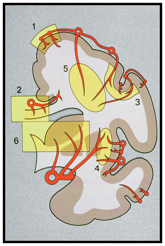
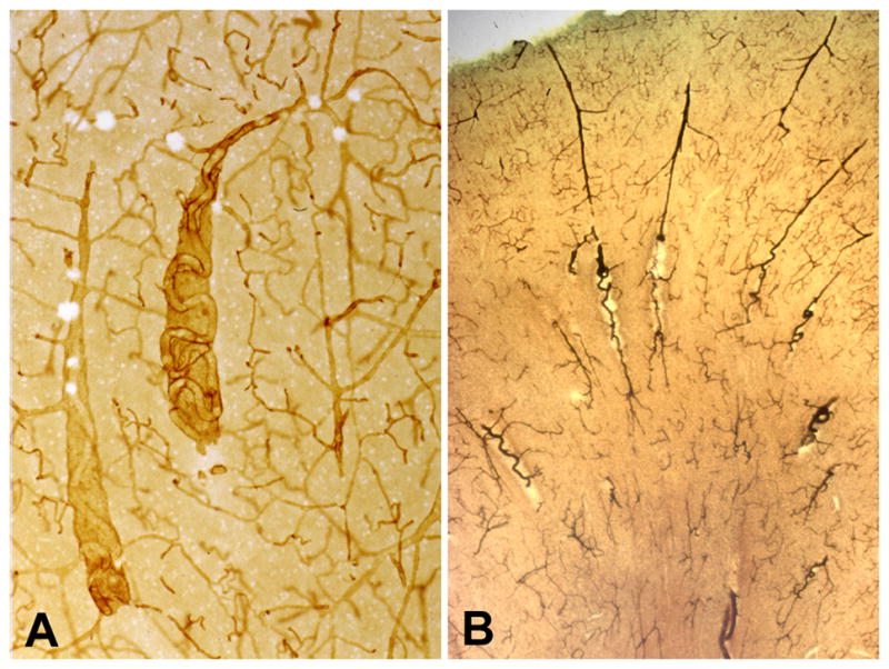
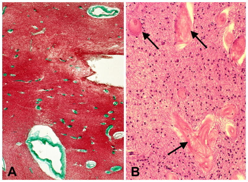
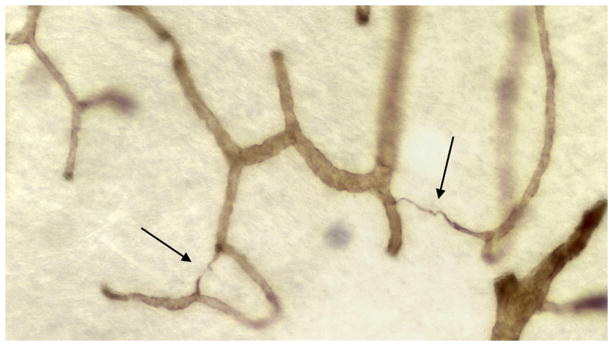
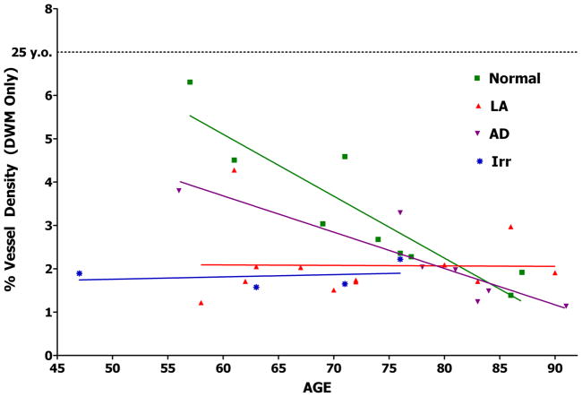
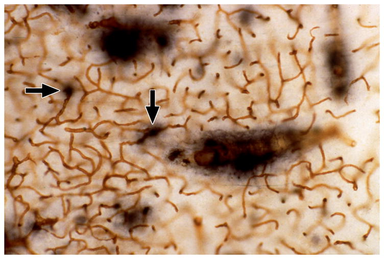
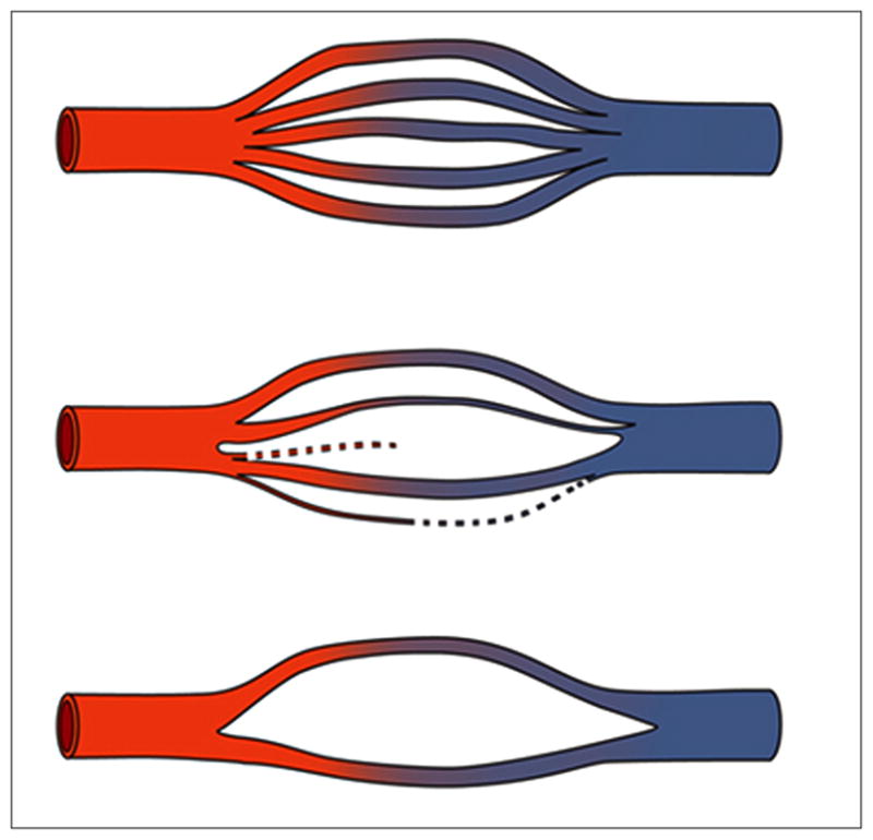
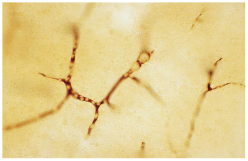
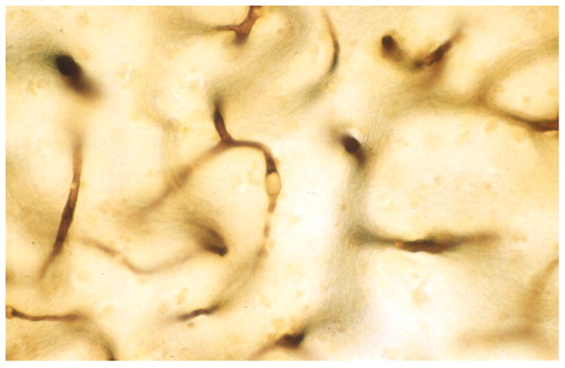
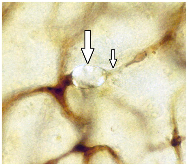
This review of age-related brain microvascular pathologies focuses on topics studied by this laboratory, including anatomy of the blood supply, tortuous vessels, venous collagenosis, capillary remnants, vascular density and microembolic brain injury. Our studies feature thick sections, large blocks embedded in celloidin, and vascular staining by alkaline phosphatase. This permits study of the vascular network in three dimensions, and the differentiation of afferent from efferent vessels. Current evidence suggests that there is decreased vascular density in ageing, Alzheimer's disease and leukoaraiosis, and cerebrovascular dysfunction precedes and accompanies cognitive dysfunction and neurodegeneration. A decline in cerebrovascular angiogenesis may inhibit recovery from hypoxia-induced capillary loss. Cerebral blood flow is inhibited by tortuous arterioles and deposition of excessive collagen in veins and venules. Misery perfusion due to capillary loss appears to occur before cell loss in leukoaraiosis, and cerebral blood flow is also reduced in the normal-appearing white matter. Hypoperfusion occurs early in Alzheimer's disease, inducing white matter lesions and correlating with dementia. In vascular dementia, cholinergic reductions are correlated with cognitive impairment, and cholinesterase inhibitors have some benefit. Most lipid microemboli from cardiac surgery pass through the brain in a few days, but some remain for weeks. They can cause what appears to be a type of vascular dementia years after surgery. Donepezil has shown some benefit. Emboli, such as clots, cholesterol crystals and microspheres can be extruded through the walls of cerebral vessels, but there is no evidence yet that lipid emboli undergo such extravasation.
© 2011 The Authors. Neuropathology and Applied Neurobiology © 2011 British Neuropathological Society.
Figures
References
-
- van Dijk EJ, Prins ND, Vrooman HA, Hofman A, Koudstaal PJ, Breteler MM. Progression of cerebral small vessel disease in relation to risk factors and cognitive consequences: Rotterdam Scan study. Stroke. 2008;39:2712–2719. - PubMed
-
- Pantoni L, Garcia JH. Pathogenesis of leukoaraiosis: a review. Stroke. 1997;28:652–659. - PubMed
-
- Hassan A, Hunt BJ, O’Sullivan M, et al. Markers of endothelial dysfunction in lacunar infarction and ischaemic leukoaraiosis. Brain. 2003;126:424–432. - PubMed
Publication types
MeSH terms
Substances
Grants and funding
LinkOut - more resources
Full Text Sources
Medical
