Impaired sodium levels in the suprachiasmatic nucleus are associated with the formation of cardiovascular deficiency in sleep-deprived rats
- PMID: 20946541
- PMCID: PMC3039182
- DOI: 10.1111/j.1469-7580.2010.01312.x
Impaired sodium levels in the suprachiasmatic nucleus are associated with the formation of cardiovascular deficiency in sleep-deprived rats
Abstract
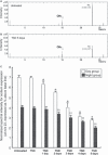
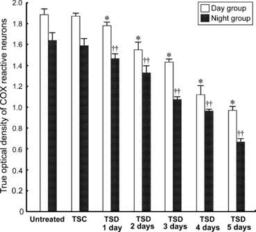
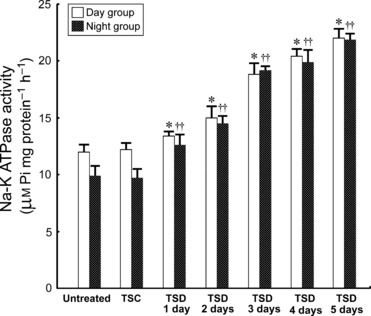
Biological rhythms are a ubiquitous feature of all higher organisms. The rhythmic center of mammals is located in the suprachiasmatic nucleus (SCN), which projects to a number of brainstem centers to exert diurnal control over many physiological processes, including cardiovascular regulation. Total sleep deprivation (TSD) is a harmful condition known to impair cardiovascular activity, but the molecular mechanisms are unknown. As the inward sodium current has long been suggested as playing an important role in driving the spontaneous firing of the SCN, the present study aimed to determine if changes in sodium expression, together with its molecular machinery (Na-K ATPase) and rhythmic activity within the SCN, would occur during TSD. Adult rats subjected to different periods of TSD were processed for time-of-flight secondary ion mass spectrometry, Na-K ATPase assay, and cytochrome oxidase (COX) (an endogenous bioenergetic marker for neuronal activity) histochemistry. Cardiovascular dysfunction was determined through analysis of heart rate and changes in mean arterial pressure. Results indicated that, in normal rats, strong sodium signals were expressed throughout the entire SCN. Enzymatic data corresponded well with spectrometric findings in which high levels of Na-K ATPase and COX were observed in this nucleus. However, following TSD, all parameters including sodium imaging, sodium intensity as well as COX activities were drastically decreased. Na-K ATPase showed an increase in responsiveness following TSD. Both heart rate and mean arterial pressure measurements indicated an exaggerated pressor effect following TSD treatment. As proper sodium levels are essential for SCN activation, reduced SCN sodium levels may interrupt the oscillatory control, which could serve as the underlying mechanism for the initiation or development of TSD-related cardiovascular deficiency.
© 2010 The Authors. Journal of Anatomy © 2010 Anatomical Society of Great Britain and Ireland.
Figures


Similar articles
-
Total sleep deprivation inhibits the neuronal nitric oxide synthase and cytochrome oxidase reactivities in the nodose ganglion of adult rats.J Anat. 2006 Aug;209(2):239-50. doi: 10.1111/j.1469-7580.2006.00594.x. J Anat. 2006. PMID: 16879602 Free PMC article.
-
Melatonin preserves longevity protein (sirtuin 1) expression in the hippocampus of total sleep-deprived rats.J Pineal Res. 2009 Oct;47(3):211-20. doi: 10.1111/j.1600-079X.2009.00704.x. Epub 2009 Jul 21. J Pineal Res. 2009. PMID: 19627456
-
Sleep deprivation impairs Ca2+ expression in the hippocampus: ionic imaging analysis for cognitive deficiency with TOF-SIMS.Microsc Microanal. 2012 Jun;18(3):425-35. doi: 10.1017/S1431927612000086. Epub 2012 Apr 12. Microsc Microanal. 2012. PMID: 22494489
-
Cardiovascular control by the suprachiasmatic nucleus: neural and neuroendocrine mechanisms in human and rat.Biol Chem. 2003 May;384(5):697-709. doi: 10.1515/BC.2003.078. Biol Chem. 2003. PMID: 12817466 Review.
-
Ion Channels Controlling Circadian Rhythms in Suprachiasmatic Nucleus Excitability.Physiol Rev. 2020 Oct 1;100(4):1415-1454. doi: 10.1152/physrev.00027.2019. Epub 2020 Mar 12. Physiol Rev. 2020. PMID: 32163720 Free PMC article. Review.
Cited by
-
Plasmon-activated water effectively relieves hepatic oxidative damage resulting from chronic sleep deprivation.RSC Adv. 2018 Mar 5;8(18):9618-9626. doi: 10.1039/c7ra13559a. eCollection 2018 Mar 5. RSC Adv. 2018. PMID: 35540828 Free PMC article.
References
-
- Adret P, Margoliash D. Metabolic and neural activity in the song system nucleus robustus archistriatalis: effect of age and gender. J Comp Neurol. 2002;454:409–423. - PubMed
-
- Adya HVA, Mallick BN. Comparison of Na-K ATPase in the rat brain synaptosome under different conditions. Neurochem Int. 1998;33:283–286. - PubMed
-
- Belle MDC, Diekman CO, Forger DB, et al. Daily electrical silencing in the mammalian circadian clock. Science. 2009;326:281–284. - PubMed
-
- Belu AM, Graham DJ, Castner DG. Time-of-flight secondary ion mass spectrometry: techniques and applications for the characterization of biomaterial surfaces. Biomaterials. 2003;24:3635–3653. - PubMed
Publication types
MeSH terms
Substances
LinkOut - more resources
Full Text Sources
Research Materials
Miscellaneous

