

P-wave indices: derivation of reference values from the Framingham Heart Study
- PMID: 20946557
- PMCID: PMC3394095
- DOI: 10.1111/j.1542-474X.2010.00390.x
P-wave indices: derivation of reference values from the Framingham Heart Study
Abstract
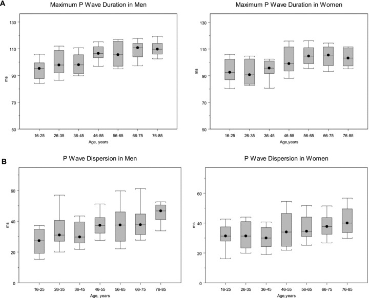
Background: P-wave indices, an electrocardiographic phenotype reflecting atrial electrophysiology and morphology, may be altered in multiple disease states or by cardiovascular risk factors. Reference values for P-wave indices, providing cut points for their classification and interpretation, have not yet been established and are essential toward facilitating clinical application and comparison between studies.
Methods: We randomly selected 20 men and 20 women from 10-year age intervals between <25 years to 76-85 years from the Framingham Heart Study Original and Offspring Cohorts, excluding subjects with prevalent cardiovascular disease, hypertension, diabetes or obesity. The total included 295 subjects; eligibility in women >75 years was limited by exclusion criteria. We used a digital measurement technique with demonstrated intrarater reproducibility to determine P-wave indices. P-wave indices examined included the maximum, mean, lead II and PR durations, dispersion, and the standard deviation of duration.
Results: All P-wave indices were significantly (P < 0.0001) correlated with advancing age. Means of all P-wave indices were lower in women as compared to men. PR-interval duration was strongly correlated with maximum, mean, and lead II mean P-wave durations. In multivariable models adjusting for significant anthropometric and clinical associations risk factors, significant differences persisted by age and sex in P-wave indices.
Conclusions: In our healthy sample without cardiovascular disease, hypertension, diabetes, or obesity, men and older subjects had longer mean P-wave indices. Our description of P-wave indices establishes reference values for future comparative studies and facilitates the classification of P-wave indices.
©2010, Wiley Periodicals, Inc.
Figures
Similar articles
-
P-wave indices, distribution and quality control assessment (from the Framingham Heart Study).Ann Noninvasive Electrocardiol. 2010 Jan;15(1):77-84. doi: 10.1111/j.1542-474X.2009.00343.x. Ann Noninvasive Electrocardiol. 2010. PMID: 20146786 Free PMC article.
-
Reference ranges of PR duration and P-wave indices in individuals free of cardiovascular disease: the Multi-Ethnic Study of Atherosclerosis (MESA).J Electrocardiol. 2013 Nov-Dec;46(6):702-6. doi: 10.1016/j.jelectrocard.2013.05.006. Epub 2013 Jun 24. J Electrocardiol. 2013. PMID: 23806475 Free PMC article.
-
P wave indices, obesity, and the metabolic syndrome: the atherosclerosis risk in communities study.Obesity (Silver Spring). 2012 Mar;20(3):666-72. doi: 10.1038/oby.2011.53. Epub 2011 Apr 7. Obesity (Silver Spring). 2012. PMID: 21475136 Free PMC article.
-
Short-term repeatability of electrocardiographic P wave indices and PR interval.J Electrocardiol. 2014 Mar-Apr;47(2):257-63. doi: 10.1016/j.jelectrocard.2013.11.007. Epub 2013 Nov 25. J Electrocardiol. 2014. PMID: 24360345 Free PMC article.
-
P wave analysis indices in young healthy men: data from the digital electrocardiographic study in Hellenic Air Force Servicemen (DEHAS).Pacing Clin Electrophysiol. 2003 Jan;26(1P2):367-72. doi: 10.1046/j.1460-9592.2003.00051.x. Pacing Clin Electrophysiol. 2003. PMID: 12687847
Cited by
-
Automatically assessed P-wave predicts cardiac events independently of left atrial enlargement in patients with cardiovascular risks: The Japan Morning Surge-Home Blood Pressure Study.J Clin Hypertens (Greenwich). 2021 Feb;23(2):301-308. doi: 10.1111/jch.14136. Epub 2020 Dec 19. J Clin Hypertens (Greenwich). 2021. PMID: 33340234 Free PMC article.
-
Meta-analysis of p-wave dispersion values in healthy individuals: the influence of clinical characteristics.Ann Noninvasive Electrocardiol. 2012 Jan;17(1):28-35. doi: 10.1111/j.1542-474X.2011.00478.x. Ann Noninvasive Electrocardiol. 2012. PMID: 22276626 Free PMC article.
-
Assessment of Hypertension Using Clinical Electrocardiogram Features: A First-Ever Review.Front Med (Lausanne). 2020 Dec 4;7:583331. doi: 10.3389/fmed.2020.583331. eCollection 2020. Front Med (Lausanne). 2020. PMID: 33344473 Free PMC article.
-
Systemic Inflammation Rapidly Induces Reversible Atrial Electrical Remodeling: The Role of Interleukin-6-Mediated Changes in Connexin Expression.J Am Heart Assoc. 2019 Aug 20;8(16):e011006. doi: 10.1161/JAHA.118.011006. Epub 2019 Aug 19. J Am Heart Assoc. 2019. PMID: 31423933 Free PMC article.
-
Assessment of atrial electromechanical interval and P wave dispersion in patients with polycystic ovary syndrome.Anatol J Cardiol. 2016 Feb;16(2):100-5. doi: 10.5152/akd.2015.5735. Epub 2015 Mar 23. Anatol J Cardiol. 2016. PMID: 26467368 Free PMC article.
References
-
- Gialafos EJ, Dilaveris PE, Synetos AG, et al P wave analysis indices in young healthy men: Data from the digital electrocardiographic study in Hellenic Air Force Servicemen (DEHAS). Pacing Clin Electrophysiol 2003;26(1 Pt 2):367–372. - PubMed
-
- Asad N, Spodick DH. Prevalence of interatrial block in a general hospital population. Am J Cardiol 2003;91:609–610. - PubMed
Publication types
MeSH terms
Grants and funding
LinkOut - more resources
Full Text Sources
Research Materials
