. 2009:468:239-58.
doi: 10.1016/S0076-6879(09)68012-5.

Structural analysis of RNA in living cells by in vivo synchrotron X-ray footprinting

Affiliations

Structural analysis of RNA in living cells by in vivo synchrotron X-ray footprinting

Tadepalli Adilakshmi et al. Methods Enzymol. 2009.

Abstract

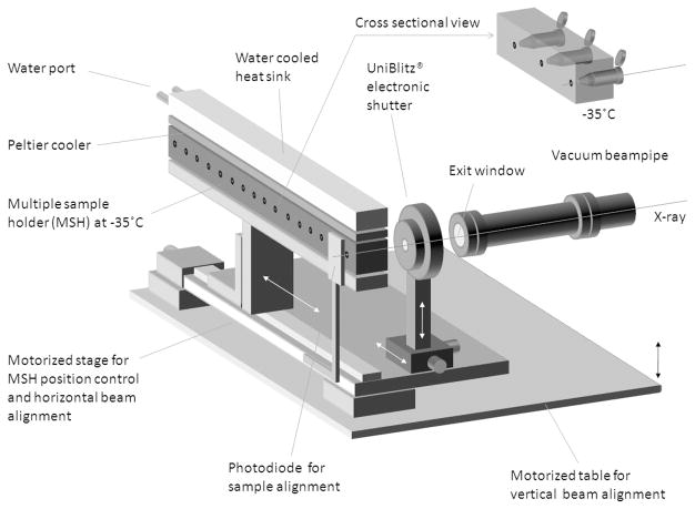
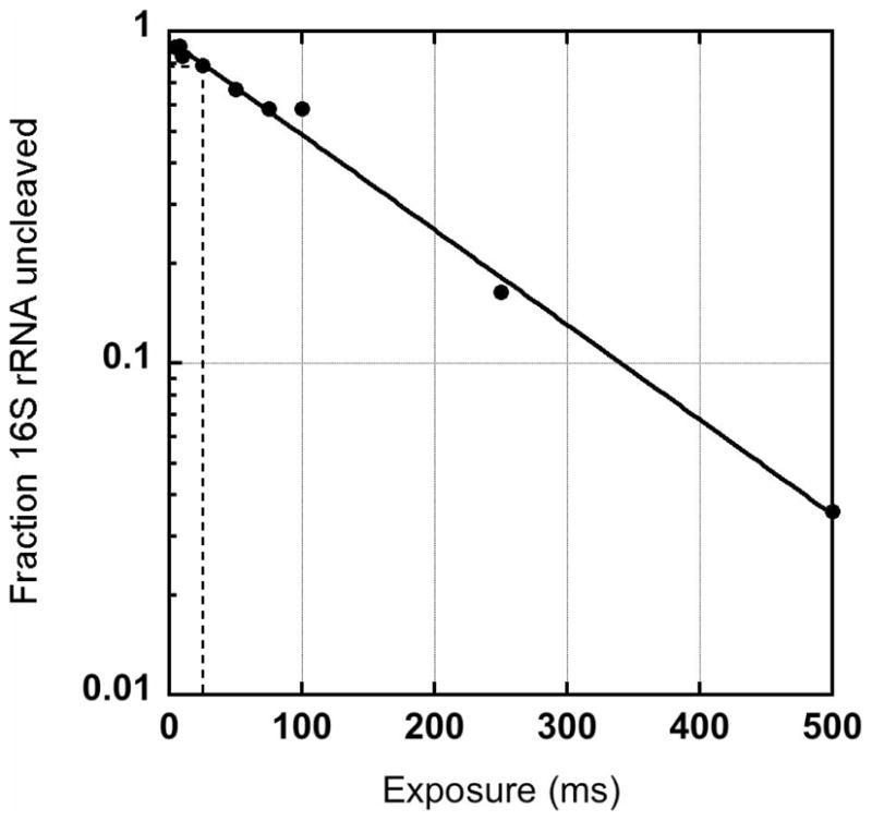
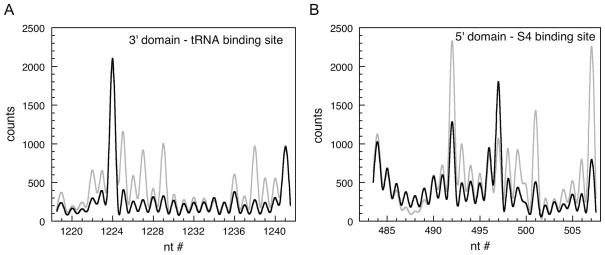
Chemical footprinting methods are widely used to probe the solution structures of nucleic acids and their complexes. Among the many available modifying reagents, hydroxyl radical is exceptional in its ability to provide nucleotide-level information on the solvent accessibility of the nucleic acid backbone. Until recently, hydroxyl radical footprinting has been limited to in vitro experiments. We describe the use of synchrotron X-radiation to generate hydroxyl radicals within cells for effective footprinting of RNA-protein complexes in vivo. This technique gives results that are consistent with in vitro footprinting experiments, with differences reflecting apparent structural changes to the RNA in vivo.


Figures

Figure 1
Figure 1
Multiple sample holder for irradiation of cell pellets. Block holding 23 samples can be cooled to −38 ºC was custom fabricated for use at beamline X28C. Samples are automatically aligned with the X-ray beam using a photodiode and motorized table. Exposure times are gated by an electronic shutter. Figure courtesy of S. Gupta, Brookhaven National Laboratory.
Figure 2
Figure 2
Determining the correct X-ray dose for footprinting. Dose-response curve showing relative fraction of intact 16S rRNA versus time of X-ray exposure of intact, frozen E. coli cells. The optimal dose in this experiment is obtained after 25–50 ms exposure. The relative amount of intact rRNA was determined by primer extension as described in the text.
Figure 3
Figure 3
X-ray footprinting of ribosomes in intact E. coli. Primer extension of purified 70S ribosomes (A) or rRNA isolated from irradiated cells (B). Exposure times are given at the top of the gel. Radiolabeled primer anneals after nt 560 of the 16S rRNA. Product bands were assigned by comparison with dideoxy sequencing lanes. Redrawn from (Adilakshmi et al. 2006).
Figure 4
Figure 4
Solvent accessibility of rRNA by hydroxyl radical footprinting. Sequencing gels in Figure 3 were quantified using a Phosphorimager. In this region of the gel, peaks due to individual nucleotides are well-resolved. Black, 70S ribosomes (in vitro); red, E. coli cells (in vivo). (A) Region from 16S 3′ domain. (B) Region from 16S 5′ domain. Reprinted from (Adilakshmi et al. 2006).

Similar articles

Cited by

References

    1. Adilakshmi T, Lease RA, Woodson SA. Hydroxyl radical footprinting in vivo: mapping macromolecular structures with synchrotron radiation. Nucleic Acids Res. 2006;34(8):e64. - PMC - PubMed
    1. Balzer M, Wagner R. A chemical modification method for the structural analysis of RNA and RNA-protein complexes within living cells. Anal Biochem. 1998;256(2):240–242. - PubMed
    1. Burkhoff AM, Tullius TD. The unusual conformation adopted by the adenine tracts in kinetoplast DNA. Cell. 1987;48(6):935–943. - PubMed
    1. Cadet J, Bellon S, Douki T, Frelon S, Gasparutto D, Muller E, Pouget JP, Ravanat JL, Romieu A, Sauvaigo S. Radiation-induced DNA damage: formation, measurement, and biochemical features. J Environ Pathol Toxicol Oncol. 2004;23(1):33–43. - PubMed
    1. Cassler MR, Grimwade JE, McGarry KC, Mott RT, Leonard AC. Drunken-cell footprints: nuclease treatment of ethanol-permeabilized bacteria reveals an initiation-like nucleoprotein complex in stationary phase replication origins. Nucleic Acids Res. 1999;27(23):4570–4576. - PMC - PubMed

Publication types