. 2010 Oct 15;143(2):263-74.
doi: 10.1016/j.cell.2010.09.022.

Patronin regulates the microtubule network by protecting microtubule minus ends

Affiliations

Patronin regulates the microtubule network by protecting microtubule minus ends

Sarah S Goodwin et al. Cell. .

Abstract

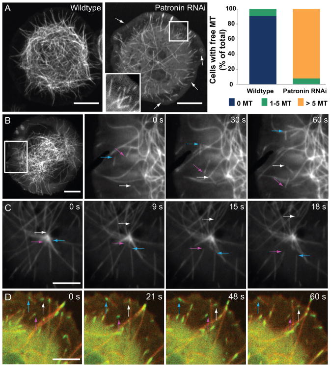
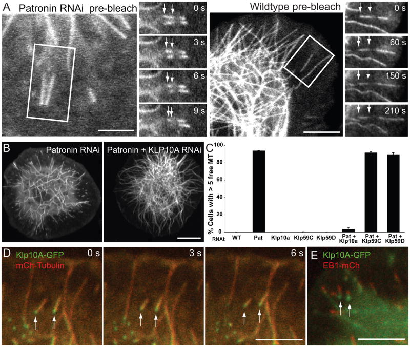
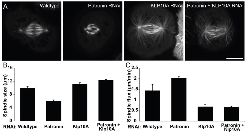
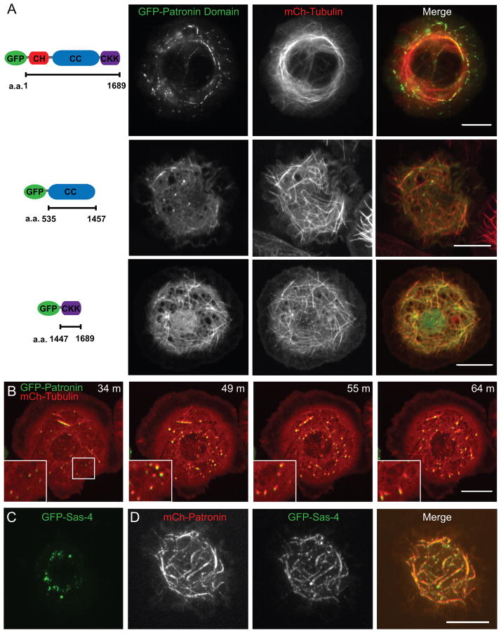
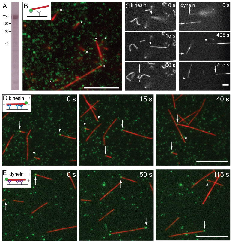
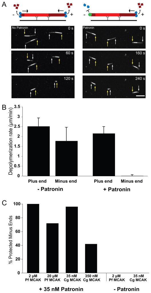
Tubulin assembles into microtubule polymers that have distinct plus and minus ends. Most microtubule plus ends in living cells are dynamic; the transitions between growth and shrinkage are regulated by assembly-promoting and destabilizing proteins. In contrast, minus ends are generally not dynamic, suggesting their stabilization by some unknown protein. Here, we have identified Patronin (also known as ssp4) as a protein that stabilizes microtubule minus ends in Drosophila S2 cells. In the absence of Patronin, minus ends lose subunits through the actions of the Kinesin-13 microtubule depolymerase, leading to a sparse interphase microtubule array and short, disorganized mitotic spindles. In vitro, the selective binding of purified Patronin to microtubule minus ends is sufficient to protect them against Kinesin-13-induced depolymerization. We propose that Patronin caps and stabilizes microtubule minus ends, an activity that serves a critical role in the organization of the microtubule cytoskeleton.


Figures

Figure 1
Figure 1. Depletion of Patronin results in free microtubules that move through the cytoplasm
Time-lapse microscopy of wildtype and Patronin-depleted GFP-tubulin Drosophila S2 cells. (A) Patronin-depleted cells have numerous free microtubules (both the plus and minus end of the same microtubule are clearly visible, arrows) which are rarely seen in wildtype cells and also have a sparser microtubule network (insert shows a region with several free microtubules). The right side shows the quantitation of free microtubules per cell from two independent experiments; colored bars indicate the percentage of cells with the number of indicated free microtubules observed (n = 200 cells per experiment; SEM <6%). Scale bars, 10 μm. See Movie S1. (B) Time-lapse TIRF microscopy of Patronin-depleted GFP-tubulin cells demonstrates that free microtubules move throughout the cytoplasm (colored arrows follow the motion of the leading end of three microtubules). Scale bar, 10 μm. See Movie S2. (C) In Patronin-depleted cells, microtubules (arrows) release and move away from the centrosome (prophase cell). Scale bar, 5 μm. See Movie S2. (D) In cells co-expressing EB1-GFP (green) and mCherry-tubulin (red), EB1 localizes to the leading end of moving microtubules (arrows), indicating that this is the microtubule plus end. See Movie S3. Brightness was adjusted in each color channel separately. Scale bar, 5 μm. See also Figures S1 and S2.
Figure 2
Figure 2. Free microtubules move by Klp10A-mediated treadmilling in Patronin-depleted cells
(A) Photobleaching a mark in the middle of moving microtubules in Patronin RNAi cells reveal that the bleach mark is stationary and the trailing minus end moves towards the bleach mark (see arrows)(n=20). This indicates that the apparent motion of microtubules occurs through simultaneous tubulin polymerization at the plus end and depolymerization at the minus end. In wildtype cells, the bleach mark in a rare free microtubule remains stationary relative to the minus end, indicating that it is neither polymerizing nor depolymerizing (n=10). Scale bars, 10 μm. (B) Comparison of GFP-tubulin cells depleted of Patronin alone or both Patronin and Klp10A. Cells co-depleted of Patronin and Klp10A have a wildtype-like microtubule network and rarely have free microtubules. Scale bar, 10 μm. (C) Quantitation of the percentage of cells with >5 free microtubules shows that co-depletion of Patronin and Klp10A, but not Klp59C or Klp59D, rescues the Patronin RNAi phenotype. The mean and SEM is shown from two independent experiments (n=200 cells per experiment). (D) In Patronin-depleted cells co-expressing Klp10A-GFP (green) and mCherry-tubulin (red), Klp10A localizes to and tracks along the depolymerizing minus end of treadmilling microtubules (arrows). Scale bar, 5 μm. See Movie S3. (E) In Patronin-depleted cells co-expressing Klp10A-GFP (green) and EB1-mCherry (red), Klp10A localizes to the trailing end, while EB1 localizes to the leading end of treadmilling free microtubules (frame from a time-lapse sequence). Scale bar, 5 μm. Brightness was adjusted in each color channel separately. See also Figures S1 and S2.
Figure 3
Figure 3. Depletion of Klp10A suppresses the Patronin phenotype in mitosis
(A) Co-depletion of Patronin and Klp10A rescues the short spindle phenotype observed in Patronin-depleted cells and results in elongated spindles similar to those seen in Klp10A-depleted cells. Scale bar, 10 μm. (B) The mean pole-to-pole metaphase spindle length under each condition was quantified for two independent experiments (n>60 spindles per condition; error bar, SEM; p<0.001 for each reported condition). (C) The flux of tubulin towards the spindle poles was measured by photobleaching a ~1 μm stripe in the GFP-tubulin spindle and tracking its movement. The mean flux rates were quantified under each condition from two independent experiments (n=20 spindles per condition; error bar, SEM; p< 0.001 for each reported condition except the pair of Klp10A RNAi and Klp10A/Patronin RNAi flux (p<0.9)). Thus poleward flux is increased after Patronin depletion and decreased below wildtype levels when Patronin and Klp10A are co-depleted. See also Figure S3.
Figure 4
Figure 4. GFP-Patronin localization and domain analysis
(A) Co-expression GFP-fusions of full-length Patronin (TIRF microscopy) or Patronin domains with mCherry-tubulin (merge: GFP-Patronin in green and mCherry-tubulin in red). Localization patterns are discussed in the text. Scale bars, 10 μm. (B) Time-lapse microscopy of GFP-Patronin (green) and mCherry-tubulin (red) expressing cells re-growing their microtubule network after washout of the microtubule depolymerizing drug colcemid (time after washout is indicated). The inserts correspond to the box at 34 min. Patronin and tubulin localize to small foci, which serve as points of microtubule nucleation during the reformation of the cytoskeleton. (C) Cells expressing GFP-Sas-4 alone form cytoplasmic foci, but when GFP-Sas-4 is co-expressed with mCherry-Patronin (D), Sas-4 is recruited to sites of mCherry-Patronin along microtubules. Brightness was adjusted in each color channel separately in the merged images. See also Figures S4 and S5.
Figure 5
Figure 5. Purified Patronin selectively binds to microtubule minus ends in vitro
(A) Purified GFP-Patronin-6xHis analyzed by SDS polyacrylamide gel electrophoresis and stained with Coomassie blue. Immunoblot analysis reveals that lower band of the doublet is Patronin lacking the GFP (not shown). (B) When GFP-Patronin is attached to a coverslip with anti-GFP antibody, it binds GMP-CPP stabilized rhodamine-labeled microtubules by one end (see Movie S4). Asterisks indicate the site of microtubule anchoring, which often overlaps with a GFP-Patronin spot. Scale bar, 10 μm. (C) To reveal which microtubule end was anchored to GFP-Patronin, kinesin or dynein was added after microtubule anchoring. Arrows follow a microtubule that was initially anchored by one end and then bound along its length to the motor-covered surface. With kinesin, the formerly anchored end is leading moving end (until the leading end reattaches and the microtubule buckles (asterisk, 60 s); with dynein, the formerly anchored end is trailing. See Movie S5. These assays reveal that microtubules are anchored to surface-bound Patronin selectively at their minus end (see statistics from three independent experiments in the text). Scale bar, 5 μm. Conventional kinesin (D) or dynein (E) microtubule gliding assays in the presence of GFP-Patronin (6 nM; green) demonstrate that GFP-Patronin binds selectively to the minus end. In the kinesin assay, GFP-Patronin (green) is most frequently observed at the leading end of gliding microtubules, while in the dynein assay, it resides at the trailing end. The results from three independent experiments indicate that GFP-Patronin binds selectively to the minus end. See Movie S6. Scale bars, 10 μm. Brightness was adjusted in each color channel separately.
Figure 6
Figure 6. GFP-Patronin protects microtubule minus ends from Kinesin-13-induced depolymerization in vitro
(A) Polarity marked GMP-CPP-stabilized rhodamine-labeled microtubules were attached to the coverslip by an anti-rhodamine antibody. The minus end is closest to the region of higher fluorescence intensity in the microtubule. In the absence of Patronin, purified Kinesin-13 motor domain from P. falciparum (3 μM) depolymerizes both ends of the microtubule. In contrast, in the presence of GFP-Patronin (30 nM), Kinesin-13 only depolymerizes the dimmer, plus end (white arrows) while the minus end (yellow arrows) is stable. See Movie S7. (Note: the higher concentration of Patronin precludes imaging of individual Patronins at microtubule ends as in Fig. 5). Scale bar, 10 μm. (B) Quantitation of Kinesin-13-induced depolymerization rates at the plus and minus ends (n = 30 microtubules for each condition; mean and SD). Data is representative of three independent experiments with different microtubule preparations. (C) Patronin was mixed with the indicated concentration of either full-length Kinesin-13 from hamster (C.g.) or the motor domain from P. falciparum (P.f.) and added to polarity marked microtubules. Minus ends that showed no detectable depolymerization by the time the plus end depolymerized by >50% of the microtubule length was scored as protected. Higher concentrations of the motors are able to compete with Patronin to depolymerize a subset of minus ends. Percentages are representative of two independent experiments. See also Figure S6.

Comment in

  • Cytoskeleton: Keeping minus ends stable.
    Wrighton KH. Wrighton KH. Nat Rev Mol Cell Biol. 2010 Dec;11(12):816. doi: 10.1038/nrm3014. Epub 2010 Nov 11. Nat Rev Mol Cell Biol. 2010. PMID: 21068769 No abstract available.

References

    1. Akhmanova A, Steinmetz M. Tracking the ends: a dynamic protein network controls the fate of microtubule tips. Nat Rev Mol Cell Biol. 2008;9:309–22. - PubMed
    1. Baines AJ, Bignone PA, King MD, Maggs AM, Bennett PM, Pinder JC, Phillips GW. The CKK domain (DUF1781) domain binds microtubules and defines the CAMSAP/ssp4 family of animal proteins. Mol Biol Evol. 2009;26:2005–14. - PubMed
    1. Bartolini F, Gundersen GG. Generation of noncentrosomal microtubule arrays. J Cell Sci. 2006;119:4155–63. - PubMed
    1. Berglund L, Björling E, Oksvold P, Fagerberg L, Asplund A, Al-Khalili Szigyarto C, Persson A, Ottosson J, Wernérus H, Nilsson P, et al. A genecentric Human Protein Atlas for expression profiles based on antibodies. Mol Cell Proteomics. 2008;7:2019–27. - PubMed
    1. Bornens M. Centrosome composition and microtubule anchoring mechanisms. Curr Opin Cell Biol. 2002;14:25–34. - PubMed

Publication types

MeSH terms