. 2010 Oct 13;18(10):1300-10.
doi: 10.1016/j.str.2010.07.008.

Cryo-EM analysis reveals new insights into the mechanism of action of pyruvate carboxylase

Affiliations

Cryo-EM analysis reveals new insights into the mechanism of action of pyruvate carboxylase

Gorka Lasso et al. Structure. .

Abstract

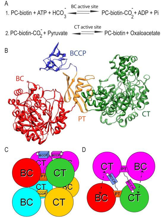
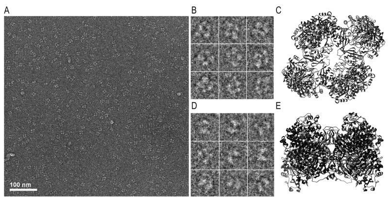
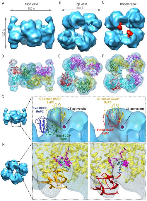
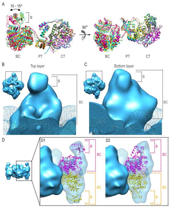
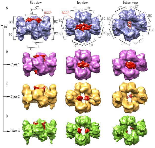
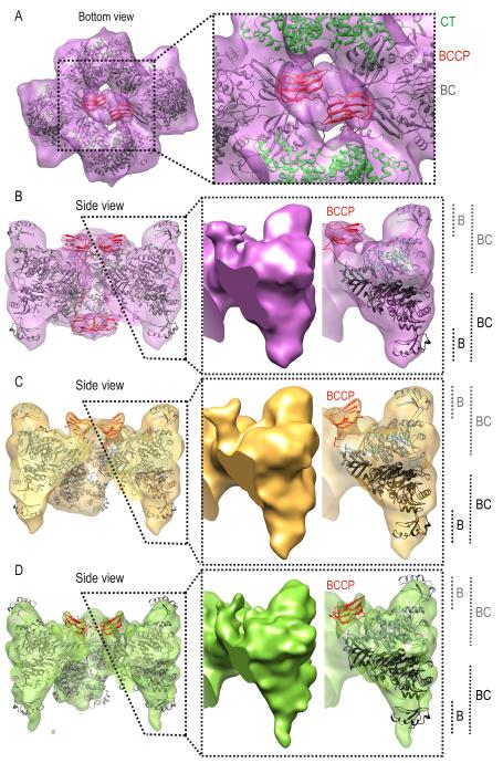
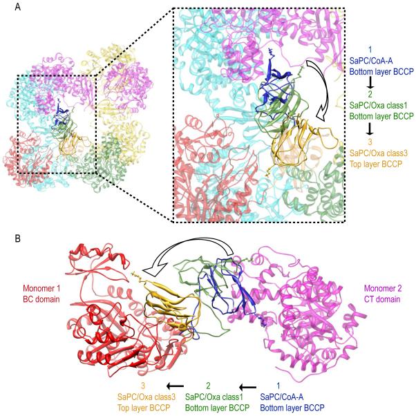
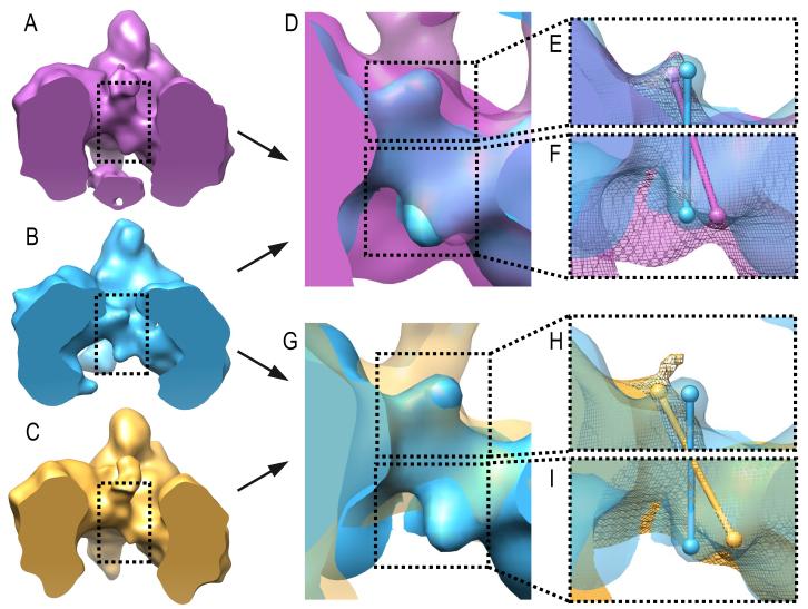
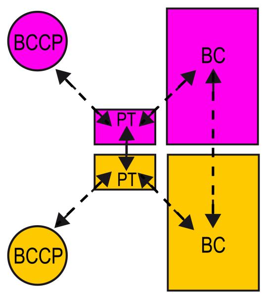
Pyruvate carboxylase (PC) is a conserved multifunctional enzyme linked to important metabolic diseases. PC homotetramer is arranged in two layers with two opposing monomers per layer. Cryo-EM explores the conformational variability of PC in the presence of different substrates. The results demonstrate that the biotin-carboxyl carrier protein (BCCP) domain localizes near the biotin carboxylase (BC) domain of its own monomer and travels to the carboxyltransferase (CT) domain of the opposite monomer. All density maps show noticeable conformational differences between layers, mainly for the BCCP and BC domains. This asymmetry may be indicative of a coordination mechanism where monomers from different layers catalyze the BC and CT reactions consecutively. A conformational change of the PC tetramerization (PT) domain suggests a new functional role in communication. A long-range communication pathway between subunits in different layers, via interacting PT-PT and BC-BC domains, may be responsible for the cooperativity of PC from Staphylococcus aureus.


Figures

Figure 1
Figure 1
Schema of the pyruvate carboxylase arrangement and function. (A) Chemical reactions carried out by PC. (B) Domain composition of the pyruvate carboxylase monomer: the biotin carboxylase (BC) domain in red, the protein tetramerization (PT) domain in orange, the carboxyl transferase (CT) domain in green and the biotin carboxyl carrier protein (BCCP) in blue. (C) A side view of the two layers of dimers comprising the homotetramer. (D) Model for the sequential chemical reactions: the biotin linked to the BCCP (in white) is carboxylated in the BC domain of its own monomer, and the transfer of the carboxyl group to pyruvate produces oxaloacetate in the CT domain of the opposite monomer.
Figure 2
Figure 2
SaPC observed by cryo-EM. (A) Micrograph corresponding to SaPC upon addition of acetyl-CoA and AMPPNP. (B) Particles defined by square-like shape delimited by four rounded regions (one on each corner) and a central cavity, that resemble (C) the top view of the SaPC atomic model based on 3BG5. (D) Particles characterized by two parallel pseudo-ellipses that were joined together at a particular region and resemble (E) the lateral view of the SaPC atomic model based on 3BG5.
Figure 3
Figure 3
Electron density map of SaPC/CoA-A at σ = 3.44 and fitting of the atomic coordinates corresponding to SaPC (PDB code = 3BG5). (A) Lateral view. (B) Top view. (C) Bottom view, density colored in red corresponds to the BCCP domain. (D-F) Electron density map and the fitted atomic structure, each subunit is shown in a different color. (D) Lateral view. (E) Top view. (F) Bottom view. (G) Rigid fitting of RePC (PDB code: 2QF7) and SaPC within the electron density map. Displayed structures correspond to the exo BCCP domain of RePC (blue), exo BCCP domain of SaPC (green), CT active BCCP domain (yellow) and manually fitted BCCP domain into the obtained density map (red). (H) Close-up of the electron density region corresponding to the CT active site on the bottom layer. The atomic structure corresponding to the CT domain is shown in yellow, residues important for biotin (in blue) binding are highlighted in purple (Xiang and Tong, 2008): Ser911, Lys912, Gln575, Ala610, Arg644, Tyr651, Thr908. The BCCP domain corresponding to the 3BG5 active site position is shown in gold and the BCCP domain fitted into the obtained electron density map is shown in red.
Fig 4
Fig 4
Conformational differences of the BC domain on top and bottom layers in SaPC/CoA-A electron density map. (A) Monomer superposition of the atomic model after flexible fitting. (B) Electron density region corresponding to the BC domain on the top layer. (C) Electron density region corresponding to the BC domain on the bottom layer. (D) Fitting of the assembled model into the given electron density map with the BC domains in (D1) open and (D2) closed conformation. Monomers are shown in magenta and yellow.
Figure 5
Figure 5
Electron density map of SaPC/Oxa using all particles (in blue) and the obtained density maps after maximum likelihood based classification (class 1 in purple, class 2 in yellow and class 3 in green). The density regions corresponding to BCCP domains are shown in red.
Figure 6
Figure 6
Fitting of the SaPC atomic model within the electron density maps obtained after maximum likelihood based classification: (A-B) SaPC/Oxa class 1 at a sigma value σ of 2.48. (A) Bottom view of the SaPC/Oxa class 1 density map with the fitted atomic model, the CT and BCCP domains are highlighted in green and red respectively. (B) Lateral view of the clipped density map of SaPC/Oxa class 1 and the fitted atomic model, BCCP domains are highlighted in red. (C) Lateral view of the clipped density map of SaPC/Oxa class 2 at a sigma value σ of 3.2 and the fitted atomic model, BCCP domains are highlighted in red. (D) Lateral view of the clipped density map of SaPC/Oxa class 3 at a sigma value σ of 3.76 and the fitted atomic model, BCCP domains are highlighted in red.
Figure 7
Figure 7
BCCP transition from the CT active site of the opposite monomer (Monomer 2, in magenta) to the BC domain of its own monomer (Monomer 1, in red). BCCP transition is shown by superimposing the fitted BCCP domains in (i) the bottom layer of SaPC/CoA-A (in blue), (ii) the bottom layer of SaPC/Oxa class 1 (in green) and (iii) the top layer of SaPC/Oxa class 3 (in gold). (A) Top view. (B) Close up lateral view.
Figure 8
Figure 8
Movement of the PT domains upon addition of oxaloacetate. (A) Lateral view of the clipped electron density of SaPC/Oxa class 1 (σ = 2.48). (B) Lateral view of the clipped electron density of SaPC/CoA-A (σ = 3.44). (C) Lateral view of the clipped electron density of SaPC/Oxa class 1 (σ = 3.2). (D) Superposition of the electron density maps of SaPC/Oxa class 1 (purple, σ = 2.48) and SaPC/CoA-A (blue, σ = 3.44). (E-F) Close up showing the superposition of the electron density maps of SaPC/Oxa class 1 (purple) and SaPC/CoA-A (blue) on the PT domain located on the top layer and bottom layer respectively. Spheres correspond to the approximate position of the C-terminus of PT domains. (E) SaPC/Oxa class 1 σ = 4.17, SaPC/CoA-A σ = 3.44. (F) SaPC/Oxa class 1 σ = 2.48, SaPC/CoA-A σ = 3.44. (G) Superposition of the electron density maps of SaPC/Oxa class 2 (gold, σ = 3.2) and SaPC/CoA-A (blue, σ = 3.44). (H-I) Close up showing the superposition of the electron density maps of SaPC/Oxa class 2 (gold) and SaPC/CoA-A (blue) on the PT domain located on the top layer and bottom layer respectively. Spheres correspond to the approximate position of the corresponding C-terminal PT domain. (H) SaPC/Oxa class 2 σ = 4.05, SaPC/CoA-A σ = 3.44. (F) SaPC/Oxa class 2 σ = 2.65, SaPC/CoA-A σ = 3.44. See also Figure S6.
Figure 9
Figure 9
Schema of the communication model between domains and layers in SaPC. The dashed arrows indicate the possible communication paths between domains.

References

    1. Attwood PV. The structure and the mechanism of action of pyruvate carboxylase. Int. J. Biochem. Cell Biol. 1995;27:231–249. - PubMed
    1. Attwood PV, Cleland WW. Decarboxylation of oxalacetate by pyruvate carboxylase. Biochemistry. 1986;25:8191–8196. - PubMed
    1. Attwood PV, Johannssen W, Chapman-Smith A, Wallace JC. The existence of multiple tetrameric conformers of chicken liver pyruvate carboxylase and their roles in dilution inactivation. Biochem. J. 1993;290(Pt 2):583–590. - PMC - PubMed
    1. Baxter WT, Leith A, Frank J. SPIRE: the SPIDER reconstruction engine. J. Struct. Biol. 2007;157:56–63. - PubMed
    1. Bottcher B, Wynne SA, Crowther RA. Determination of the fold of the core protein of hepatitis B virus by electron cryomicroscopy. Nature. 1997;386:88–91. - PubMed

Publication types

MeSH terms