. 2010 Oct 13;18(10):1332-41.
doi: 10.1016/j.str.2010.07.007.

Hydrogen/deuterium exchange reveals distinct agonist/partial agonist receptor dynamics within vitamin D receptor/retinoid X receptor heterodimer

Affiliations

Hydrogen/deuterium exchange reveals distinct agonist/partial agonist receptor dynamics within vitamin D receptor/retinoid X receptor heterodimer

Jun Zhang et al. Structure. .

Abstract

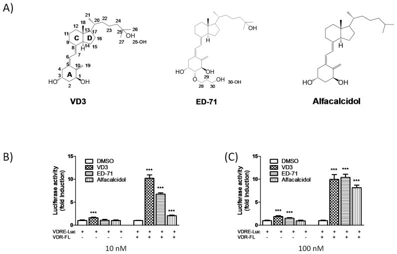
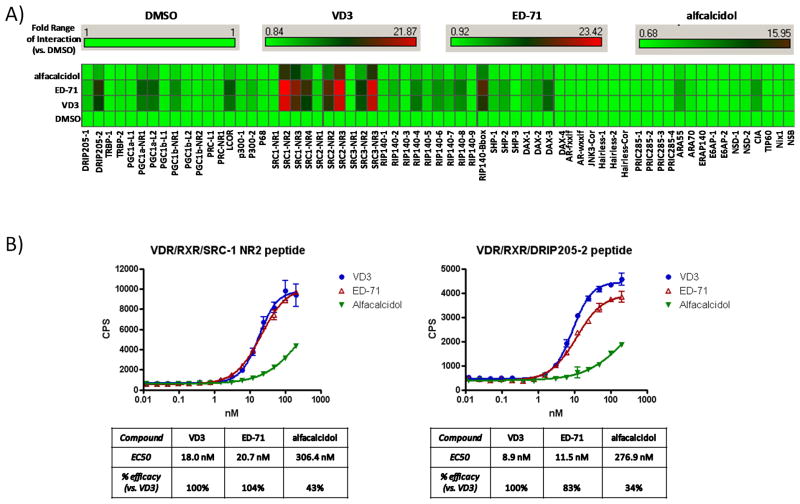
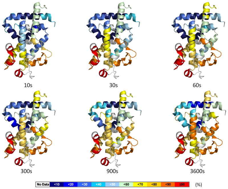
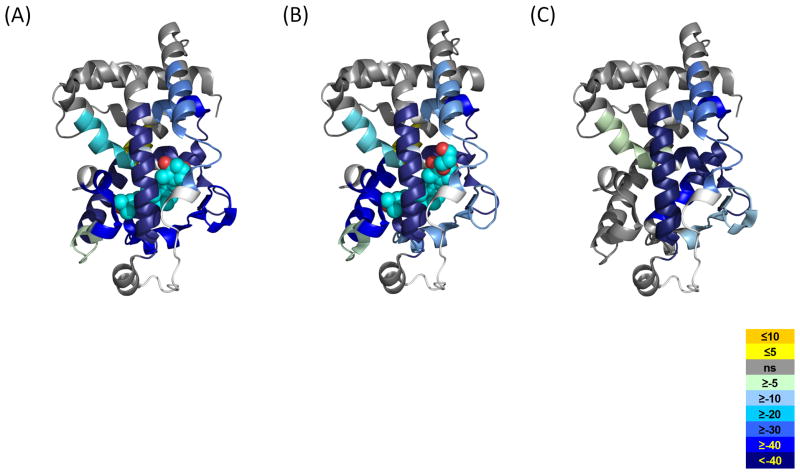
Regulation of nuclear receptor (NR) activity is driven by alterations in the conformational dynamics of the receptor upon ligand binding. Previously, we demonstrated that hydrogen/deuterium exchange (HDX) can be applied to determine novel mechanism of action of PPARγ ligands and in predicting tissue specificity of selective estrogen receptor modulators. Here, we applied HDX to probe the conformational dynamics of the ligand binding domain (LBD) of the vitamin D receptor (VDR) upon binding its natural ligand 1α,25-dihydroxyvitamin D3 (1,25D3), and two analogs, alfacalcidol and ED-71. Comparison of HDX profiles from ligands in complex with the LBD with full-length receptor bound to its cognate receptor retinoid X receptor (RXR) revealed unique receptor dynamics that could not be inferred from static crystal structures. These results demonstrate that ligands modulate the dynamics of the heterodimer interface as well as provide insight into the role of AF-2 dynamics in the action of VDR partial agonists.


Figures

Figure 1
Figure 1
VDR ligands and transcriptional responses. A) Chemical structures of the ligands. B) HEK293 T cells were transfected with the 4xVDRE reporter plasmid and VDR-FL or empty vector and treated with different VDR modulators (10 nM and 100 nM) or DMSO.
Figure 2
Figure 2
Quantitative measurement of cofactor peptide interactions. A) Full length human His-VDR and Flag-RXR proteins were added to a multiplexed streptavidin-based bead-bound cassette of biotinylated NHR cofactor peptides. VDR ligands were added to this mixture at fully saturable concentrations (1μM) and incubated with shaking for 2hrs. Quantitation of VDR/ligand/RXR/bead-bound cofactor peptide complexes were measured using xMAP-based BioRad Luminex 120 instrument and Applied Cytometry Systems StarStation software. Cofactor peptide interactions with VDR/RXR are displayed as a heat map (fold interaction vs. the Apo DMSO state) using Spotfire DecisionSuite software. B) Using anti-Flag/streptavidin bead-based alphascreen technology, full length human His-VDR and Flag-RXR proteins were incubated with either biotinylated SRC-1 NR2 or DRIP205-2 33-mer cofactor peptides. VDR ligands were added to this mixture with increasing concentration (10 nM and 100 nM) and after 15min the formation of VDR/RXR/peptide complexes were quantified using a PerkinElmer Envision 2103 Multilabel Reader. Data analysis and curve-fitting was performed using GraphPad Prizm software.
Figure 3
Figure 3
Dynamics of apo VDR-LBD. The percentage of deuterium levels for all peptides of apo VDR-LBD is mapped with color for each exchange point onto PDB:1DB1; the color code is explained at the bottom of the figure. Regions colored as white represent peptide are not detected. See also Movie S1.
Figure 4
Figure 4
Average differential HDX of each ligand mapped onto corresponding VDR-LBD/ligand crystal structures. A) 1,25D3 (PDB:1DB1), B) ED-71 (PDB:2HAR), C) Alfacalcidol (PDB:1DB1, since there is no VDR-LBD/alfacalcidol crystal structure available). The color legend shows the differential HDX between Apo VDR-LBD and ligand-bound ones. The regions in the crystal structure colored as white represent where are not covered in this study. See also Figures S1 and S3.

Comment in

References

    1. Berrodin TJ, Chang KC, Komm BS, Freedman LP, Nagpal S. Differential biochemical and cellular actions of Premarin estrogens: distinct pharmacology of bazedoxifene-conjugated estrogens combination. Mol Endocrinol. 2009;23:74–85. - PMC - PubMed
    1. Bruning JB, Chalmers MJ, Prasad S, Busby SA, Kamenecka TM, He Y, Nettles KW, Griffin PR. Partial agonists activate PPARgamma using a helix 12 independent mechanism. Structure. 2007;15:1258–1271. - PubMed
    1. Chalmers MJ, Busby SA, Pascal BD, He Y, Hendrickson CL, Marshall AG, Griffin PR. Probing protein ligand interactions by automated hydrogen/deuterium exchange mass spectrometry. Anal Chem. 2006;78:1005–1014. - PubMed
    1. Chalmers MJ, Busby SA, Pascal BD, Southern MR, Griffin PR. A two-stage differential hydrogen deuterium exchange method for the rapid characterization of protein/ligand interactions. J Biomol Tech. 2007;18:194–204. - PMC - PubMed
    1. Chandra V, Huang P, Hamuro Y, Raghuram S, Wang Y, Burris TP, Rastinejad F. Structure of the intact PPAR-gamma-RXR-alpha nuclear receptor complex on DNA. Nature. 2008:350–356. - PMC - PubMed

Publication types

MeSH terms

LinkOut - more resources