. 2010 Dec 31;285(53):41961-71.
doi: 10.1074/jbc.M110.169607. Epub 2010 Oct 14.

Identification of microRNAs regulating reprogramming factor LIN28 in embryonic stem cells and cancer cells

Affiliations

Identification of microRNAs regulating reprogramming factor LIN28 in embryonic stem cells and cancer cells

Xiaomin Zhong et al. J Biol Chem. .

Abstract

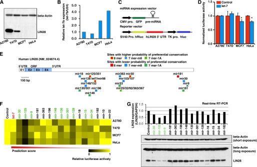
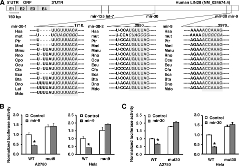
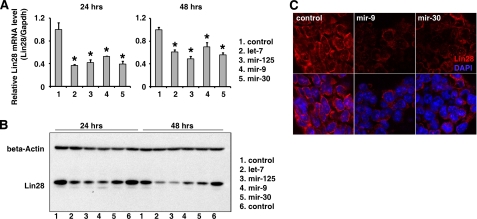
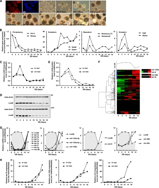
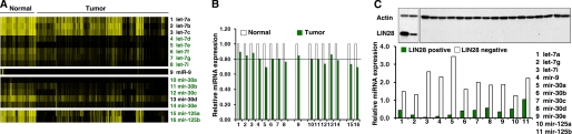
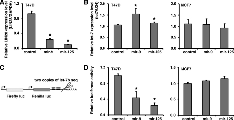
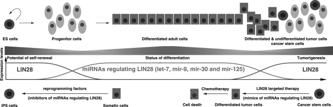
LIN28 (a homologue of the Caenorhabditis elegans lin-28 gene) is an evolutionarily conserved RNA-binding protein and a master regulator controlling the pluripotency of embryonic stem cells. Together with OCT4, SOX2, and NANOG, LIN28 can reprogram somatic cells, producing induced pluripotent stem cells. Expression of LIN28 is highly restricted to embryonic stem cells and developing tissues. In human tumors, LIN28 is up-regulated and functions as an oncogene promoting malignant transformation and tumor progression. However, the mechanisms of transcriptional and post-transcriptional regulation of LIN28 are still largely unknown. To examine microRNAs (miRNAs) that repress LIN28 expression, a combined in silico prediction and miRNA library screening approach was used in the present study. Four miRNAs directly regulating LIN28 (let-7, mir-125, mir-9, and mir-30) were initially identified by this approach and further validated by quantitative RT-PCR, Western blot analysis, and a LIN28 3'-UTR reporter assay. We found that expression levels of these four miRNAs were clustered together and inversely correlated with LIN28 expression during embryonic stem cell differentiation. In addition, the expression of these miRNAs was remarkably lower in LIN28-positive tumor cells compared with LIN28-negative tumor cells. Importantly, we demonstrated that these miRNAs were able to regulate the expression and activity of let-7, mediated by LIN28. Taken together, our studies demonstrate that miRNAs let-7, mir-125, mir-9, and mir-30 directly repress LIN28 expression in embryonic stem and cancer cells. Global down-regulation of these miRNAs may be one of the mechanisms of LIN28 reactivation in human cancers.


Figures

FIGURE 1.
FIGURE 1.
A combined in silico prediction and miRNA library screening approach identified miRNAs targeting LIN28. A, Western blots were used to detect endogenous LIN28 expression in the four cancer cell lines selected for experimental screening. Two of these (A2780 and T47D) were LIN28-positive, and two (MCF7 and HeLa) were LIN28-negative. B, real-time RT-PCR was used to detect let-7b expression in these cell lines. As expected, the LIN28-negative lines expressed relatively higher levels of let-7b. C, illustration of the miRNA expression vector and reporter vector used in the screening assay. Pro., promoter; hRluc, Renilla luciferase; hluc, firefly luciferase. D, the known LIN28-regulatory miRNA let-7 was used for the pilot screening. Co-transfection with the let-7b expression vector significantly (*, p < 0.05) reduced the luciferase activity of the LIN28 3′-UTR reporter vector in the two LIN28-negative cell lines. E, schematic structure of LIN28 mRNA. The miRNA binding sites were predicted by TargetScan. UTR, untranslated region; ORF, open reading frame; E, exon. F, the summary heat map of the miRNA library screening in four cell lines. Here, miRNAs are listed from left to right according to their prediction scores (high to low). Nine miRNAs (marked in green) significantly reduced the reporter activity in all four cancer cell lines. G, stable cell lines overexpressing each of these nine miRNAs as well as five miRNA controls that did not reduce luciferase activity were generated by lentiviral infection. Three of the nine candidate miRNAs (mir-30, mir-125, and mir-9) markedly reduced both LIN28 mRNA and protein expression.
FIGURE 2.
FIGURE 2.
LIN28-regulatory function of mir-9 and mir-30 was validated by the 3′-UTR reporter assay. A, schematic diagram of the mir-125, let-7, mir-30, and mir-9 binding sites in the LIN28 3′-UTR. The seeding sequences (marked in gray) were broadly conserved among different species. Hsa, Human; Ptr, chimpanzee; Mml, Rhesus; Mmu, mouse; Rno, Rat; Cpo, Pig; Ocu, rabbit; Eeu, Hedgehog; Cfa, Dog; Eca, Horse; Bta, Cow; Dno, armadillo; Laf, elephant; Mdo, opossum. B and C, summary of the reporter assays on the wild type and mir-9 binding site mutant reporter (B) and mir-30 binding site mutant reporter (C) in A2780 (LIN28-positive) and HeLa (LIN28-negative) cells. WT, wild type LIN28 3′-UTR reporter; mut9, mir-9 binding site mutant LIN28 3′-UTR reporter; mut30, mir-30 binding site mutant LIN28 3′-UTR reporter. Overexpression of mir-9 or mir-30 was able to significantly (*, p < 0.05) reduce luciferase activity in the wild type but not the binding site mutant LIN28 3′-UTR reporters.
FIGURE 3.
FIGURE 3.
miRNAs regulate Lin28 in undifferentiated ES cells. The miRNA mimics (30 nm) of let-7, mir-125, mir-9, mir-30, and a control mimic were transiently transfected into the mouse ES cell line R1. At 24 and 48 h post-transfection, total RNA and protein were collected, and the endogenous Lin28 expression was examined by real-time RT-PCR (A) and Western blots (B). A, the Lin28 mRNA expression was significantly (*, p < 0.05) decreased in the cells transfected with miRNA mimics compared with the control transfections. B, Lin28 protein expression was markedly decreased in the cells transfected with miRNA mimics compared with the control transfections. C, immunohistochemical staining further confirmed that mir-9 and mir-30 decreased LIN28 expression in ES cells.
FIGURE 4.
FIGURE 4.
Expression levels of Lin28 and its regulatory miRNAs are inversely correlated during ES cell differentiation. A, ES cells were differentiated in vitro by spontaneously self-assembling in suspension culture into three-dimensional cell aggregates (EBs). Immunostaining demonstrated that Lin28 was highly expressed in the undifferentiated ES cells. B, pluripotency and differentiation markers during differentiation and EB formation were monitored by real-time RT-PCR. C, Lin28 mRNA expression during EB formation was analyzed by real-time RT-PCR. D and E, Lin28 protein expression during EB formation was analyzed using Western blots. F, the global miRNA expression profile during EB formation was analyzed using a TaqMan miRNA assay. An unsupervised cluster analysis indicated that all four Lin28-regulatory miRNAs grouped together. G, detailed miRNA expression changes during EB formation identified by the TaqMan miRNA assay. All four Lin28-regulatory miRNAs were markedly up-regulated from day 6 of EB formation. H, real-time RT-PCR validations of the TaqMan miRNA assay in two ES cell lines.
FIGURE 5.
FIGURE 5.
LIN28-regulatory miRNAs are globally down-regulated in LIN28-positive cancer cell lines. A, a publicly available miRNA microarray data set was retrieved from the Broad Institute (66). Normalized expression levels of miRNAs regulating LIN28 were analyzed and are shown as a heat map. We found that 11 of 16 LIN28-regulatory miRNAs (marked in green) were markedly down-regulated (more than 20% reduction) in human tumors compared with normal control tissues. B, summary of the average expression levels of the miRNAs regulating LIN28 in normal and tumor specimens in the public miRNA microarray data set. C, LIN28 protein levels in 19 human cancer cell lines detected by Western blot. Two of these cell lines (10.5%) were LIN28-positive. The LIN28-regulatory miRNA expression levels were analyzed by real-time RT-PCR in these 19 cell lines. Shown is a summary of the average expression of each individual miRNA in the LIN28-positive lines (green) and LIN28-negative lines (white).
FIGURE 6.
FIGURE 6.
miRNAs regulate let-7 expression mediated by LIN28. A, the transfection of mir-9 and mir-125 mimics significantly (*, p < 0.05) reduced LIN28 mRNA expression in T47D cells. B, transfection of mir-9 and mir-125 mimics significantly (*, p < 0.05) increased let-7b expression in the LIN28-positive cell line T47D but not in the LIN28-negative cell line MCF7. C, a miRNA-responsive sensor, a technique for monitoring miRNA activity, bearing sequences complementary to let-7b in the downstream region of the 3′-UTR of a constitutively expressed reporter gene. D, transfection of mir-9 and mir-125 mimics significantly (*, p < 0.05) decreased the let-7b-responsive sensor in the LIN28-positive cell line T47D but not in the LIN28-negative cell line MCF7.
FIGURE 7.
FIGURE 7.
The regulatory circuitry afforded by LIN28-regulatory miRNAs in development and tumorigenesis.

Similar articles

Cited by

References

    1. Ambros V., Horvitz H. R. (1984) Science 226, 409–416 - PubMed
    1. Moss E. G., Lee R. C., Ambros V. (1997) Cell 88, 637–646 - PubMed
    1. Yokoyama S., Hashimoto M., Shimizu H., Ueno-Kudoh H., Uchibe K., Kimura I., Asahara H. (2008) Gene Expr. Patterns 8, 155–160 - PMC - PubMed
    1. Polesskaya A., Cuvellier S., Naguibneva I., Duquet A., Moss E. G., Harel-Bellan A. (2007) Genes Dev. 21, 1125–1138 - PMC - PubMed
    1. West J. A., Viswanathan S. R., Yabuuchi A., Cunniff K., Takeuchi A., Park I. H., Sero J. E., Zhu H., Perez-Atayde A., Frazier A. L., Surani M. A., Daley G. Q. (2009) Nature 460, 909–913 - PMC - PubMed

Publication types