. 2011 Jan;300(1):G21-32.
doi: 10.1152/ajpgi.00363.2010. Epub 2010 Oct 14.

Differential organization of excitatory and inhibitory synapses within the rat dorsal vagal complex

Affiliations

Differential organization of excitatory and inhibitory synapses within the rat dorsal vagal complex

Tanja Babic et al. Am J Physiol Gastrointest Liver Physiol. 2011 Jan.

Abstract

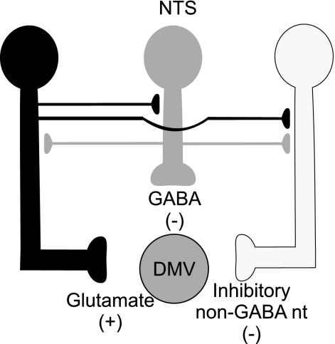
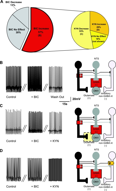
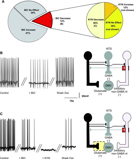
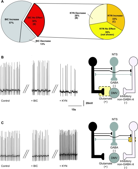
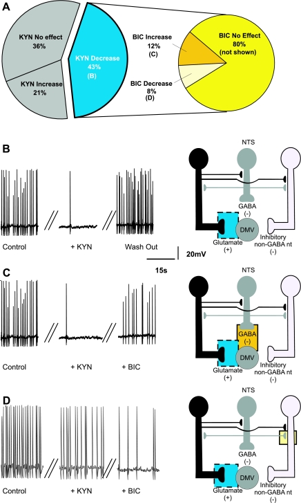
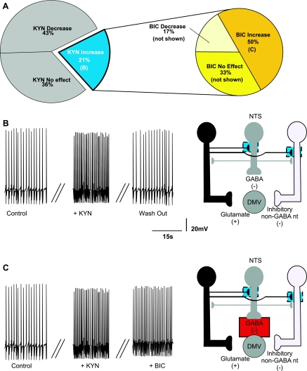
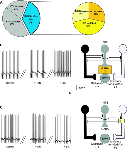
The dorsal motor nucleus of the vagus (DMV) is pivotal in the regulation of upper gastrointestinal functions, including motility and both gastric and pancreatic secretion. DMV neurons receive robust GABA- and glutamatergic inputs. Microinjection of the GABA(A) antagonist bicuculline (BIC) into the DMV increases pancreatic secretion and gastric motility, whereas the glutamatergic antagonist kynurenic acid (KYN) is ineffective unless preceded by microinjection of BIC. We used whole cell patch-clamp recordings with the aim of unveiling the brain stem neurocircuitry that uses tonic GABA- and glutamatergic synapses to control the activity of DMV neurons in a brain stem slice preparation. Perfusion with BIC altered the firing frequency of 71% of DMV neurons, increasing firing frequency in 80% of the responsive neurons and decreasing firing frequency in 20%. Addition of KYN to the perfusate either decreased (52%) or increased (25%) the firing frequency of BIC-sensitive neurons. When KYN was applied first, the firing rate was decreased in 43% and increased in 21% of the neurons; further perfusion with BIC had no additional effect in the majority of neurons. Our results indicate that there are several permutations in the arrangements of GABA- and glutamatergic inputs controlling the activity of DMV neurons. Our data support the concept of brain stem neuronal circuitry that may be wired in a finely tuned organ- or function-specific manner that permits precise and discrete modulation of the vagal motor output to the gastrointestinal tract.


Figures

Fig. 1.
Fig. 1.
Schematic diagram showing glutamate (black), GABA (gray), and inhibitory non-GABAergic (white) neurons impinging on the dorsal motor nucleus of the vagus (DMV). Note that we do not mean to imply that the synaptic contacts occur specifically on the dendrites, axons, or synaptic terminals, but they are depicted in this manner for schematic clarity. NTS, nucleus tractus solitarius.
Fig. 2.
Fig. 2.
Pie charts (A), representative traces (BD, left), and the proposed neuronal circuits (BD, right) in neurons in which perfusion with bicuculline (BIC) increased the firing rate of DMV neurons. Perfusion with BIC increased the firing frequency of DMV neurons (N = 35), and perfusion with kynurenic acid (KYN) decreased the firing frequency of the majority of these neurons. Note that the pie chart on the right represents subgroups of neurons in which BIC increased the firing frequency. The increase in firing rate induced by BIC is likely due to antagonism of GABAA receptors located either on the membrane of the DMV neuron or on tonically active glutamatergic neurons that impinge on the DMV neuron. Perfusion with KYN in the presence of BIC decreased the firing rate of DMV neurons (N = 19). This response was likely due to KYN-mediated inhibition of a tonic glutamatergic input onto DMV neurons. Perfusion with KYN in the presence of BIC further increased the firing rate of DMV neurons (N = 10), suggesting the presence of tonic glutamatergic synapses impinging onto non-GABAergic neurons. In all figures, the proposed site of action of bicuculline is indicated by a solid-line red square and the proposed site of action of kynurenic acid is indicated by a dotted-line yellow square.
Fig. 3.
Fig. 3.
Pie charts (A), representative traces (B and C, left), and the proposed neuronal circuits (B and C, right) in neurons in which perfusion with BIC decreased the firing rate of DMV neurons. Perfusion with BIC decreased the firing frequency of DMV neurons (N = 9), and KYN further decreased the firing frequency in majority of these neurons. The pie chart on the right represents subgroups of neurons in which BIC decreased the firing frequency. The decrease in firing rate induced by BIC is likely due to antagonism of GABAA receptors located on the membrane of non-GABAergic inhibitory neurons. Perfusion with KYN in the presence of BIC further decreased the firing rate of DMV neurons (N = 4). This response was likely due to KYN-mediated inhibition of a tonic glutamatergic input onto DMV neurons.
Fig. 4.
Fig. 4.
Pie charts (A), representative traces (B and C, left), and the proposed neuronal circuits (B and C, right) in neurons in which perfusion with BIC did not affect the firing rate (N = 18) of DMV neurons. A: perfusion with KYN in the presence of BIC decreased the firing rate of DMV neurons (N = 5). B: the decrease in the firing rate in response to KYN was likely due to KYN-mediated inhibition of a tonic glutamatergic input onto DMV neurons. C: perfusion with KYN in the presence of BIC increased the firing rate of DMV neurons (N = 6), suggesting the presence of tonic glutamatergic synapses impinging onto non-GABAergic neurons.
Fig. 5.
Fig. 5.
Pie charts (A), representative traces (BD, left), and the proposed neuronal circuits (BD, right) in neurons in which perfusion with KYN decreased the firing rate of DMV neurons. A: perfusion with KYN decreased (or abolished) the firing frequency of DMV neurons (N = 25), and BIC had no additional effect on the firing frequency in majority of these neurons. B: the decrease in firing rate induced by KYN is likely due to antagonism of glutamatergic receptors located on the membrane of the DMV neuron. C: perfusion with BIC in the presence of KYN increased the firing rate of DMV neurons (N = 3). This response was likely due to BIC-mediated inhibition of a tonic GABAergic input onto DMV neurons. D: perfusion with BIC in the presence of KYN further decreased the firing rate of DMV neurons (N = 2), suggesting the presence of tonic GABAergic synapses impinging onto non-GABAergic neurons.
Fig. 6.
Fig. 6.
Pie charts (A), representative traces (B and C, left), and the proposed neuronal circuits (B and C, right) in neurons in which perfusion with KYN increased the firing rate of DMV neurons. A: perfusion with KYN increased the firing frequency of DMV neurons (N = 12) and perfusion with BIC further altered the firing frequency of 8 of these neurons. B: the increase in firing rate induced by KYN is likely determined by antagonism of tonically active glutamatergic receptors located on the membrane of either GABAergic or inhibitory non-GABAergic neurons. C: perfusion with BIC in the presence of KYN further increased the firing rate of DMV neurons (N = 6). This response was likely due to a BIC-mediated inhibition of a tonic GABAergic input onto DMV neurons.
Fig. 7.
Fig. 7.
Pie charts (A), representative traces (B and C, left), and the proposed neuronal circuits (B and C, right) in neurons in which perfusion with KYN did not affect the firing rate (N = 21) of DMV neurons. Perfusion with BIC in the presence of KYN either increased (N = 6) (B) or decreased (N = 6) (C) the firing rate of DMV neurons (N = 6). The increase in the firing frequency was likely due to BIC-mediated inhibition of a tonic GABAergic input onto DMV neurons. Perfusion with BIC in the presence of KYN decreased the firing rate of DMV neurons (N = 6), suggesting the presence of tonic GABAergic synapses impinging onto non-GABAergic neurons.

References

    1. Altschuler SM, Bao X, Bieger D, Hopkins DA, Miselis RR. Viscerotopic representation of the upper alimentary tract in the rat: sensory ganglia and nuclei of the solitary and spinal trigeminal tracts. J Comp Neurol 283: 248–268, 1989 - PubMed
    1. Andresen MC, Yang M. Non-NMDA receptors mediate sensory afferent synaptic transmission in medial nucleus tractus solitarius. Am J Physiol Heart Circ Physiol 259: H1307–H1311, 1990 - PubMed
    1. Aylwin ML, Horowitz JM, Bonham AC. NMDA receptors contribute to primary visceral afferent transmission in the nucleus of the solitary tract. J Neurophysiol 77: 2539–2548, 1997 - PubMed
    1. Bai D, Zhu G, Pennefather P, Jackson MF, MacDonald JF, Orser BA. Distinct functional and pharmacological properties of tonic and quantal inhibitory postsynaptic currents mediated by gamma-aminobutyric acid(A) receptors in hippocampal neurons. Mol Pharmacol 59: 814–824, 2001 - PubMed
    1. Bailey TW, Appleyard SM, Jin YH, Andresen MC. Organization and properties of GABAergic neurons in solitary tract nucleus (NTS). J Neurophysiol 99: 1712–1722, 2008 - PubMed

Publication types

MeSH terms

LinkOut - more resources