. 2010 Oct 15;330(6002):385-90.
doi: 10.1126/science.1188472.

Cell type-specific loss of BDNF signaling mimics optogenetic control of cocaine reward

Affiliations

Cell type-specific loss of BDNF signaling mimics optogenetic control of cocaine reward

Mary Kay Lobo et al. Science. .

Abstract

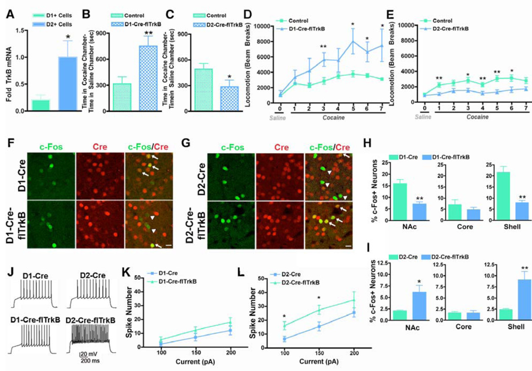
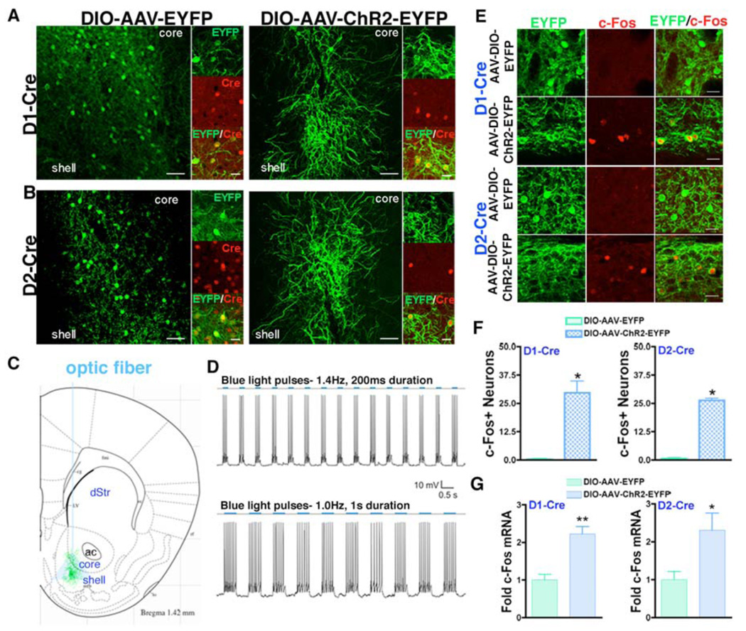
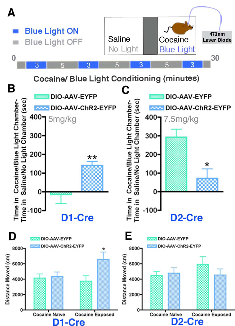
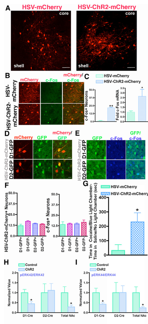
The nucleus accumbens is a key mediator of cocaine reward, but the distinct roles of the two subpopulations of nucleus accumbens projection neurons, those expressing dopamine D1 versus D2 receptors, are poorly understood. We show that deletion of TrkB, the brain-derived neurotrophic factor (BDNF) receptor, selectively from D1+ or D2+ neurons oppositely affects cocaine reward. Because loss of TrkB in D2+ neurons increases their neuronal excitability, we next used optogenetic tools to control selectively the firing rate of D1+ and D2+ nucleus accumbens neurons and studied consequent effects on cocaine reward. Activation of D2+ neurons, mimicking the loss of TrkB, suppresses cocaine reward, with opposite effects induced by activation of D1+ neurons. These results provide insight into the molecular control of D1+ and D2+ neuronal activity as well as the circuit-level contribution of these cell types to cocaine reward.


Figures

Fig 1
Fig 1. Effect of selective deletion of TrkB from D1+ or D2+ MSNs on behavioral effects of cocaine, c-Fos induction, and neuronal excitability
(A) TrkB mRNA is expressed in D1+ and D2+ MSNs FACS-purified from D1-GFP and D2-GFP transgenic mice, but is significantly enriched in D2+ MSNs (n = 4 per group); Student’s t-test, p < 0.05). (B) D1-Cre–flTrkB (n = 9) mice displayed enhanced cocaine conditioned place preference (CPP) relative to littermate controls (n = 10), whereas (C) D2-Cre–flTrkB mice (n = 14) exhibited decreased cocaine CPP compared to littermate controls (n = 16) (cocaine dose: 7.5 mg/kg ip; Student’s t-test, **p < 0.01, *p < 0.05). (D,E) D1-Cre–flTrkB and D2-Cre–flTrkB mice and littermate controls were treated with saline on day 0 and with cocaine (10 mg/kg) on days 1–7 and locomotor activity was assessed over a 30-min time period. (D) D1-Cre–flTrkB mice (n = 6) displayed enhanced cocaine-induced locomotor activity after repeated cocaine administration compared to littermate controls (n = 7) (Repeated Measures Two-Way ANOVA, genotype effect: F(1,11) = 6.20, p < 0.05; day effect: F(6,66) = 5.50, p < 0.01), while (E) D2-Cre–flTrkB mice (n = 10) showed decreased locomotor activity to acute and repeated cocaine relative to controls (n = 14) (Repeated Measures Two-Way ANOVA, genotype effect: F(1,22) = 9.98, p < 0.01; day effect: F(6,132)= 4.00, p < 0.01). Post-hoc analysis reveals significant differences on specific cocaine days (Student’s t-test, **p <0.01, *p < 0.05). Data represented as mean ± SEM. (F–I) c-Fos induction was examined 90 min after acute cocaine (20 mg/kg) by double immuno-labeling of c-Fos (green) and Cre (red) in the NAc. (F,H) D1-Cre–flTrkB mice exhibited a significant decrease in double-labeled c-Fos (green) and Cre (red) neurons in the NAc after cocaine exposure compared to D1-Cre control mice and this down-regulation is specific to the NAc shell. (G,I) In contrast, D2-Cre–flTrkB mice, relative to D2-Cre controls, displayed an increase in double-labeled c-Fos and Cre neurons in the NAc, an effect also specific to the NAc shell (n = 4 per group, Student’s t-test, **p < 0.01, *p < 0.05). Images displayed are from the NAc shell. Arrows represent neurons double labeled with c-Fos and Cre. Arrowheads represent c-Fos neurons that are not Cre positive. Scale bars, 20 µm. Data represented as mean ± SEM. (J) Sample traces obtained by 200 pA current injection (holding potential at −80 mV) in NAc shell MSNs in D1-Cre-flTrkB, D2-Cre-flTrkB, and their control mice injected with DIO-AAV-EYFP into the NAc for visualization of D1+ or D2+ MSNs. (K,L) D2+ MSNs in D2-Cre-flTrkB NAc (n=3 animals), but not from D1+ MSNs in D1-Cre-flTrkB NAc (n=4), display increased cell excitability following incremental steps in current injections (100, 150, and 200 pA) compared to respective controls, D2-Cre (n=5) and D1-Cre (n=8). Two-way ANOVA, F(1,7) = 13.23, p = 0.002 (for D2+ MSNs), F(1,11) = 4.04, p = 0.054 (for D1+ MSNs). Post hoc analysis reveals significant effects for 100 and 150 pA currents in D2+ MSNs, Student’s t-test, *p<0.05.
Fig 2
Fig 2. In vivo and in vitro optogenetic control of D1+ or D2+ MSNs
(A,B) DIO-AAV-ChR2-EYFP or DIO-AAV-EYFP was injected into the NAc of D1-Cre and D2-Cre mice resulting in ChR2-EYFP or EYFP expressing neurons (green) that also express Cre (red). Scale bars, 50 µm (low power images) and 20 µm (high power images). (C) Diagram of D1+ or D2+ ChR2 expressing MSNs and blue light emission from the optic fiber. (D) Control of neuronal firing when a NAc MSN expressing DIO-AAV-ChR2-EYFP is exposed to blue light at 1.4 or 1.0 Hz. (E) c-Fos (red) expression is induced in D1+ or D2+ MSNs expressing ChR2 (green) that have been activated with 10 Hz blue light stimulation but not EYFP expressing MSNs. Scale bars, 20 µm. (F) Quantification of E shows a significant increase in c-Fos expressing ChR2 expressing D1+ and D2+ MSNs compared to EYFP expressing controls after blue light exposure (n = 3 per group, Student’s t-test, *p < 0.01). (G) c-Fos mRNA is significantly upregulated in the NAc after blue light pulses in DIO-AAV-ChR2-EYFP expressing D1-Cre and D2-Cre mice (n = 4–5 per group, Student’s t-test, **p < 0.01, *p < 0.05).
Fig 3
Fig 3. Optogenetic activation of D1+ or D2+ MSNs oppositely regulates cocaine reward
(A) Illustration of the CPP cocaine/blue light paradigm. Mice were conditioned to a cocaine/blue light chamber and a saline/no light chamber for 30 min. Blue light pulses (10 Hz) were delivered for four 3-min periods during the 30-min conditioning. (B,C) Activating D1+ MSNs in D1-Cre DIO-AAV-ChR2-EYFP mice (n = 9) during cocaine (5 mg/kg)/blue light CPP enhances cocaine reward compared to D1-Cre DIO-AAV-EYFP controls (n = 10), whereas activating D2+ MSNs in D2-Cre DIO-AAV-ChR2-EYFP mice (n = 7) during cocaine (7.5 mg/kg)/ blue light CPP attenuates cocaine reward relative to D2-Cre DIO-AAV-EYFP controls (n = 4) (Students t-test, **p < 0.01, *p < 0.05). (D,E) Optical control of D1+ and D2+ MSNs in D1-Cre and D2-Cre mice expressing DIO-AAV-ChR2-EYFP or DIO-AAV-EYFP in a cocaine naïve state and 24 hours after 6 days of repeated cocaine (15 mg/kg) results in increased locomotor activity only when D1+ MSNs are activated after cocaine exposure (Two Way ANOVA, genotype effect: F(1,22) = 4.37, p < 0.05; Student’s t-test *p<0.05)
Fig 4
Fig 4. Global optogenetic activation of NAc neurons increases the rewarding effects of cocaine and attenuates TrkB-BDNF signaling
(A) HSV-mCherry or HSV-ChR2-mCherry (red) were injected into the NAc of wildtype mice. Scale bar 50 µm. (B) Activation of ChR2 with blue light (10 Hz) stimulation induces c-Fos (green) expression in HSV-ChR2-mCherry neurons (red), but not in HSV-mCherry neurons (red), after 10 Hz blue light. Scale bar 25 µm. (C) Quantification of c-Fos positive neurons in B and c-Fos mRNA after 10 Hz blue light shows a significant increase in c-Fos in HSV-ChR2-mCherry expressing NAc compared to HSV-mCherry expressing controls (n = 3–5 per group, Student’s t-test, **p < 0.01, *p < 0.05). (D) HSV-ChR2-mCherry (red) injection into the NAc of D1-GFP and D2-GFP mice mediates transgene expression in both D1+ and D2+ MSNs (GFP, green) and (E) c-Fos (blue) is induced by light in each MSN (green). Scale bar 10 µm. (F) Quantification of D and E shows HSV-ChR2-mCherry and blue light induced c-Fos equally in D1+ and D2+ MSNs. (G) Mice expressing HSV-ChR2-mCherry (n = 9) in the NAc displayed enhanced preference for the cocaine (5 mg/kg)/blue light chamber compared to control mice expressing HSV-mCherry (n = 8) (Student’s t-test, p < 0.05). (H,I) Blue light simulation results in a significant decrease in phospho:total ERK levels (pERK42/ERK42 and pERK44/ERK44) in the NAc of D1-Cre DIO-AAV-ChR2-EYFP mice and mice expressing HSV-ChR2-mCherry relative to their controls (DIO-AAV-EYFP or HSV-mCherry) (n = 5–8 per group, Student’s t-test *p < 0.05).

Comment in

References

    1. Hyman SE, Malenka RC, Nestler EJ. Annu Rev Neurosci. 2006;29:565. - PubMed
    1. Gerfen CR. Annu Rev Neurosci. 1992;15:285. - PubMed
    1. Albin RL, Young AB, Penney JB. Trends Neurosci. 1989 Oct;12:366. - PubMed
    1. Alexander GE, DeLong MR, Strick PL. Annu Rev Neurosci. 1986;9:357. - PubMed
    1. Graybiel AM. Curr Biol. 2000;10:R509. - PubMed

Publication types

MeSH terms