

Increase in GLUT1 in smooth muscle alters vascular contractility and increases inflammation in response to vascular injury
- PMID: 20947823
- PMCID: PMC3014530
- DOI: 10.1161/ATVBAHA.110.215004
Increase in GLUT1 in smooth muscle alters vascular contractility and increases inflammation in response to vascular injury
Abstract
Objective: The goal of this study was to test the contributing role of increasing glucose uptake in vascular smooth muscle cells (VSMCs) in vascular complications and disease.
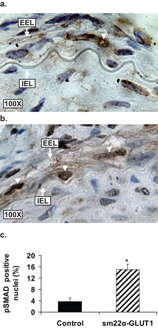
Methods and results: A murine genetic model was established in which glucose trasporter 1 (GLUT1), the non-insulin-dependent glucose transporter protein, was overexpressed in smooth muscle using the sm22α promoter. Overexpression of GLUT1 in smooth muscle led to significant increases in glucose uptake (n=3, P<0.0001) as measured using radiolabeled 2-deoxyglucose. Fasting blood glucose, insulin, and nonesterified fatty acids were unchanged. Contractility in aortic ring segments was decreased in sm22α-GLUT1 mice (n=10, P<0.04). In response to vascular injury, sm22α-GLUT1 mice exhibited a proinflammatory phenotype, including a significant increase in the percentage of neutrophils in the lesion (n=4, P<0.04) and an increase in monocyte chemoattractant protein-1 (MCP-1) immunofluorescence. Circulating haptoglobin and glutathione/total glutathione were significantly higher in the sm22α-GLUT1 mice postinjury compared with controls (n=4, P<0.05), suggesting increased flux through the pentose phosphate pathway. sm22α-GLUT1 mice exhibited significant medial hypertrophy following injury that was associated with a significant increase in the percentage of VSMCs in the media staining positive for nuclear phosphoSMAD2/3 (n=4, P<0.003).
Conclusions: In summary, these findings suggest that increased glucose uptake in VSMCs impairs vascular contractility and accelerates a proinflammatory, neutrophil-rich lesion in response to injury, as well as medial hypertrophy, which is associated with enhanced transforming growth factor-β activity.
Figures


References
-
- Intensive blood-glucose control with sulphonylureas or insulin compared with conventional treatment and risk of complications in patients with type 2 diabetes (ukpds 33). Uk prospective diabetes study (ukpds) group. Lancet. 1998;352:837–853. - PubMed
-
- Forrest KY, Becker DJ, Kuller LH, Wolfson SK, Orchard TJ. Are predictors of coronary heart disease and lower-extremity arterial disease in type 1 diabetes the same? A prospective study. Atherosclerosis. 2000;148:159–169. - PubMed
-
- Hashim S, Li Y, Anand-Srivastava MB. G protein-linked cell signaling and cardiovascular functions in diabetes/hyperglycemia. Cell Biochem Biophys. 2006;44:51–64. - PubMed
-
- Orchard TJ, Dorman JS, Maser RE, Becker DJ, Drash AL, Ellis D, LaPorte RE, Kuller LH. Prevalence of complications in iddm by sex and duration. Pittsburgh epidemiology of diabetes complications study ii. Diabetes. 1990;39:1116–1124. - PubMed
Publication types
MeSH terms
Substances
Grants and funding
LinkOut - more resources
Full Text Sources
Other Literature Sources
Molecular Biology Databases
Research Materials
Miscellaneous
