. 2011 Feb;6(2):224-35.
doi: 10.4161/epi.6.2.13700. Epub 2011 Feb 1.

Reactivation of MASPIN in non-small cell lung carcinoma (NSCLC) cells by artificial transcription factors (ATFs)

Affiliations

Reactivation of MASPIN in non-small cell lung carcinoma (NSCLC) cells by artificial transcription factors (ATFs)

Adriana S Beltran et al. Epigenetics. 2011 Feb.

Abstract

Tumor suppressor genes have antiproliferative and antimetastatic functions, and thus, they negatively affect tumor progression. Reactivating specific tumor suppressor genes would offer an important therapeutic strategy to block tumor progression. Mammary Serine Protease Inhibitor (MASPIN) is a tumor suppressor gene that is not mutated or rearranged in tumor cells, but is silenced during metastatic progression by transcriptional and epigenetic mechanisms. In this work, we have investigated the ability of Artificial Transcription Factors (ATFs) to reactivate MASPIN expression and to reduce tumor growth and metastatic dissemination in Non-Small Cell Lung Carcinoma (NSCLC) cell lines carrying a hypermethylated MASPIN promoter. We found that the ATFs linked to transactivator domains were able to demethylate the MASPIN promoter. Consistently, we observed that co-treatment of ATF-transduced cells with methyltransferase inhibitors enhanced MASPIN expression as well as induction of tumor cell apoptosis. In addition to tumor suppressive functions, restoration of endogenous MASPIN expression was accompanied by inhibition of metastatic dissemination in nude mice. ATF-mediated reactivation of MASPIN lead to changes in cell motility and to induction of E-CADHERIN. These data suggest that ATFs are able to reprogram aggressive lung tumor cells towards a more epithelial, differentiated phenotype, and thus, represent novel therapeutic agents for metastatic lung cancers.


Figures
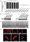
Figure 1
Figure 1
MASPIN expression in a panel of NSC LC cell lines. (A) MASPIN mRNA expression levels by quantitative real-time PCR in a panel of NSCLC cell lines. MASPIN mRNA levels were normalized to the immortalized bronchial epithelial cell line (BEAS-2B). Data represent the mean ± SD of three independent experiments. (B) MASPIN protein expression in high- and low-MASPIN expressing cell lines. (C) Loss of MASPIN expression correlates with invasion abilities in NSC LC cell lines. (D) Immunofluorescence staining of MASPIN in BEAS-2B and in the NSCLC cell lines. ↑ Indicates nuclear and ➭ indicates cytoplasmic staining, respectively.
Figure 2
Figure 2
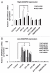
ATFs regulate MASPIN in high- and low-MASPIN expressing NSC LC cell lines. (A) Relative MASPIN mRNA upregulation in the high-MASPIN expressing cell lines NCI-H358 and NCI-H460. (B) Relative MASPIN expression in the low-MASPIN expressing cell lines NCI-A549, NCI-H157, NCI-H226, NCI-H520, NCI-H1703 and NCI-H661. MASPIN mRNA expression levels were measured by quantitative real-time PC R and data was normalized to the untransduced cell lines. Experiments were run in triplicate and data represent the mean ± SD of three independent biological replicates (*p < 0.05, as determined by ANOVA). Cell line refers to untransduced NSC LC cell lines; control, cells transduced with an empty retroviral vector; ATF-97, ATF-126 and ATF-452, cells transduced with the corresponding ATFs; ATF-126NOVP is a control ATF-126 lacking the VP64 activator domain; PMXVP64-SS is a control vector expressing the VP64 activator domain only.
Figure 3
Figure 3
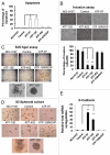
ATFs induce apoptosis and reduce cell invasion in the NCI-H157 NSCLC cell line. (A) ATF-97 and ATF-126 induce apoptosis in NCI-H157 cells. The percentage of apoptosis was quantitatively analyzed in untransduced cells, control cells and in ATF-transduced cells using an Annexin-V staining. Apoptosis was measured 96 h post-transduction. Data represents an average of three independent experiments (**p < 0.01, as determined by the Student's t-test). (B) ATFs downregulate cell invasion in NCI-H157 cells in matrigel invasion assays. Left panel shows a representative picture of invasive cells. Right panel shows the quantification of invasive cells. Experiments were run in triplicate and data represents an average of three different biological replicates (**p < 0.01, as determined by the Student's t-test). (C) In vitro colony formation assay in soft agar. The morphology of colonies of control and ATF-transduced cells are indicated. (D) 3D Matrigel culture assay illustrating that ATF-97- and ATF-126-transduced cells form colonies with an organized spheroid structure. (E) Expression of E-CADHERIN by qRT-PC R in control and ATF-transduced cells. Data was normalized to the untransduced cell line. Experiments were run in triplicate and data represent the mean ± SD three independent experiments (**p < 0.01, as determined by Student's t-test). NCI-H157 refers to untransduced cells; control, cells transduced with an empty retroviral vector; ATF-97, ATF-126 and ATF-452, cells transduced with the corresponding ATFs; ATF-126NOVP is a control ATF-126 lacking the VP64 activator domain.
Figure 4
Figure 4
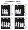
ATF-126 inhibits metastatic dissemination in the NSCLC NCI-H157 cell line stably expressing a luciferase reporter (Luc) in vivo. Top panel; in vivo Bioluminescence Imaging of control mice showing metastasis in the brain and spine at week six after receiving an intracardiac injection of 1 × 10⁵ NCI-H157 cells transduced with control vector (empty retroviral vector). Bottom panel; Bioluminescence Imaging of ATF-126-injected mice twelve weeks after receiving an intracardiac injection of 1 × 10⁵ NCI-H157 cells transduced with ATF-126 retroviral vector. Differences between the two groups were calculated using the Wilcoxon rank sum test with the level of significance of p ≤ 0.1.
Figure 5
Figure 5
ATFs demethylate the MASPIN promoter in the NCI-H157 cell line. (A) Methylation status of the MASPIN promoter in untransduced NCI-H157 cells. The X-axis represent the nucleotide position relative to the first methionine and Y-axis the percentage of methylation along the MASPIN proximal promoter region (495 to +134). The frequencies of 5-Methylcytosine were obtained by sodium bisulfite genomic sequencing of the MASPIN promoter Known transcription factor binding sites were included (p53-binding sites as well as ATF-binding sites). Red nucleotides indicate the methylated cytosines in the ATF-binding sites. (B) Schematic representation of ATF-126 and ATF-97 illustrating the positioning of the six zinc finger (ZF-97 and ZF-126) DNA binding domains and the VP64 effector domain (blue circles) in the promoter. (C) Methylation status of the MASPIN promoter in ATF-97- and ATF-126-transduced cells.
Figure 6
Figure 6
ATFs synergize with 5-Aza-2′-dC to reactivate MASPIN in NSCLC Low-MASPIN expressing cell lines. Non-transduced and transduced cells were treated with 5 µM of 5-Aza-2′-dC for 48 h. MASPIN mRNA expression levels were measured by quantitative real-time PCR. MASPIN protein expression in NCI-H157 cells was determined by western blot. Experiments were run in triplicate and data represent the mean ± SD three independent experiments (*p < 0.05, as determined by Student's t-test). NCH-A549, NCH-H226, NCH-H1703 and NCH-H157 are referred to as “cell line;” ATF-97, ATF-126 and ATF-452, represent cells transduced with the corresponding ATFs.

References

    1. Zardo G, Fazi F, Travaglini L, Nervi C. Dynamic and reversibility of heterochromatic gene silencing in human disease. Cell Res. 2005;15:679–690. - PubMed
    1. Baylin SB, Ohm JE. Epigenetic gene silencing in cancer—a mechanism for early oncogenic pathway addiction? Nat Rev Cancer. 2006;6:107–116. - PubMed
    1. Zhu S, Wu H, Wu F, Nie D, Sheng S, Mo YY. MicroRNA-21 targets tumor suppressor genes in invasion and metastasis. Cell Res. 2008;18:350–359. - PubMed
    1. Collingwood TN, Urnov FD, Wolffe AP. Nuclear receptors: coactivators, corepressors and chromatin remodeling in the control of transcription. J Mol Endocrinol. 1999;23:255–275. - PubMed
    1. Ohm JE, McGarvey KM, Yu X, Cheng L, Schuebel KE, Cope L, et al. A stem cell-like chromatin pattern may predispose tumor suppressor genes to DNA hypermethylation and heritable silencing. Nat Genet. 2007;39:237–242. - PMC - PubMed

Publication types

MeSH terms