. 2010 Oct 15;9(20):4130-43.
doi: 10.4161/cc.9.20.13389. Epub 2010 Oct 20.

p38α MAPK is a MTOC-associated protein regulating spindle assembly, spindle length and accurate chromosome segregation during mouse oocyte meiotic maturation

Affiliations

p38α MAPK is a MTOC-associated protein regulating spindle assembly, spindle length and accurate chromosome segregation during mouse oocyte meiotic maturation

Xiang-Hong Ou et al. Cell Cycle. .

Abstract

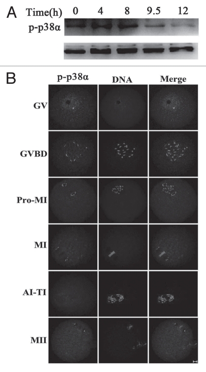
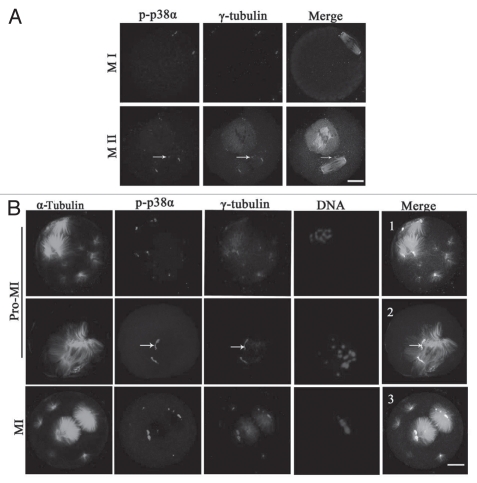
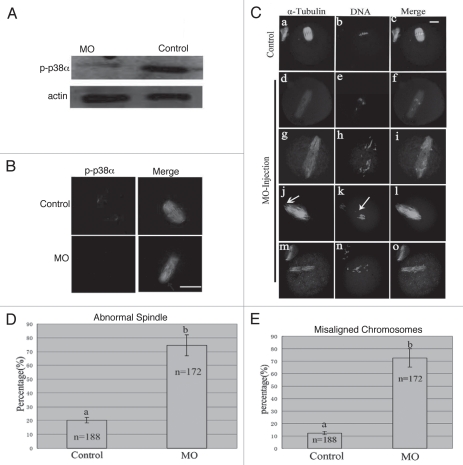
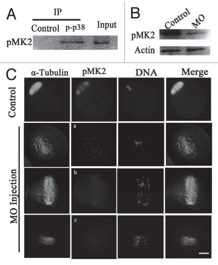
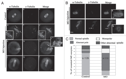
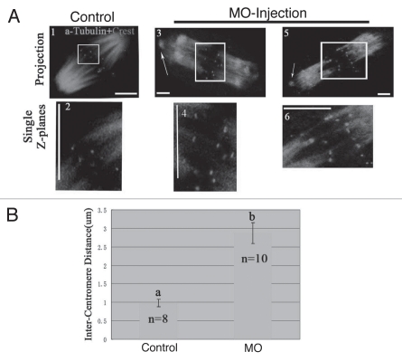
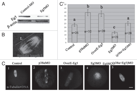
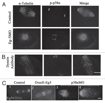
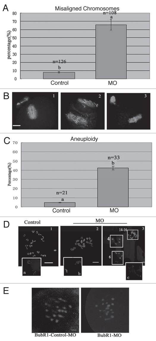
P38αMAPK (p38α) is usually activated in response to various stresses and plays a role in the inhibition of cell proliferation and tumor progression, but little is known about its roles in meiotic spindle assembly. In this study, we characterized the dynamic localization of p38α and explored its function in mouse oocyte meiotic maturation. P38α specifically colocalized with γ-tubulin and Plk1 at the center of MTOCs and spindle poles. Depletion of p38α by specific morpholino injection resulted in severely defective spindles and misaligned chromosomes probably via MK2 dephosphorylation. Notably, depletion of p38α led to significant spindle pole defects, spindle elongation, non-tethered kinetochore microtubules and increased microtubule tension. The disruption of spindle stability was coupled with decreased γ-tubulin and Plk1 at MTOCs. Overexpression of Eg5, a conserved motor protein, also caused spindle elongation and its morpholino injection almost completely rescued spindle elongation caused by p38α depletion. In addition, p38α-depletion decreased BubR1 and interfered with spindle assembly checkpoint (SAC), which resulted in aneuploid oocytes. Together, these data indicate that p38α is an important component of MTOCs, which regulates spindle assembly and spindle length, as well as stabilizes the spindle and spindle poles. Perturbed SAC and abnormal microtubule tension may be responsible for the misaligned chromosomes and high aneuploidy in p38α-depleted mouse oocytes.


Figures

Figure 1
Figure 1
Expression and subcellular localization of p-p38α during mouse oocyte meiotic maturation. (A) Expression of p-p38α and p38 during meiotic maturation at 0, 4, 8, 9.5 and 12 h, corresponding to GV, pro-MI, MI, AI-TI and MII stages, respectively. The molecular mass of p-p38α is 43 kDa and that of p38 is 43 kDa. Proteins from 300 oocytes were loaded for each sample. (B) Confocal microcopy showing immunostaining of p-p38α (green) and DNA (red) in mouse oocytes at GV (germinal vesicle), GVBD (germinal vesicle breakdown), pro-MI (first prometaphase), MI (first metaphase), AI-TI (first anaphase and telophase) and MII (second metaphase) stages. Bar = 10 µm.
Figure 2
Figure 2
p-p38α colocalizes with γ-tubulin at meiotic oocyte MTOCs and spindle poles. (A) Representative images of oocytes at MI and MII stages during meiotic maturation. p-p38 (purple) colocalization with γ-tubulin (red) was assessed, together with α-tubulin (green) and DNA (blue) staining. Arrows indicate cytoplasmic asters. Bar = 10 µm. (B) Colocalization of p-p38α and γ-tubulin at MTOCs in mouse oocytes treated with taxol. Oocytes were incubated in M2 medium containing 200 µg/ml taxol for 45 min at pro-MI and MI stages, respectively. Oocytes were stained with antibodies against p-p38α (purple), γ-tubulin (red), α-tubulin (green) and DNA (blue) dye Hoechst 33258. Arrows indicate that p-p38α is strictly colocalized with typical “C”-shaped γ-tubulin at unattached poles off spindles. Bar = 10 µm.
Figure 3
Figure 3
p38α depletion leads to defective spindles, abnormal spindle poles and misaligned chromosomes. (A) Expression of p-p38α in the p38αMO microinjected oocytes. GV oocytes were microinjected with standard control MO and p38αMO, respectively, and incubated for 24 h in M2 medium containing 2.5 µM Milrinone before oocytes were collected for western blotting. Control MO: 300 oocytes microinjected with standard control MO; MO: 300 oocytes microinjected with p38αMO. (B) Clear p-p38α signal was found at spindle poles in control MO injected oocytes; no p-p38α signal was detected in p38αMO-injected oocytes (MO). p-p38α (red); α-tubulin (green); DNA (blue). Bar = 10 µm. (C) Spindle morphology and chromosome alignment after microinjection of MO standard control and p38αMO in mouse oocytes. In the control group (n = 188), most oocytes showed normal spindle morphology and chromosome alignment (C, a and b), while in the MO injection group (n = 172), most oocytes showed severely defective spindle and chromosome alignment. Straggled (h and n) or lagging chromosomes (e and k) were found in oocytes at the MI, AI, MII stages. Spindle elongation (d, g, j and m); multiple poles (g); disintegrated poles (d, g, j and m) were observed in oocytes at MI and MII. Arrow in “k” indicates the misaligned chromosomes; Arrow in “j” shows non-tethered pole. α-tubulin (green); DNA (red). Bar = 10 µm. (D and E) Percentage of oocytes with abnormal spindles and misaligned chromosomes between the control and p38αMO groups. Data are expressed as mean ± SEM of at least three independent experiments and different letters indicate statistically significant difference (p < 0.05).
Figure 4
Figure 4
Analysis of the interaction of p38α with MK2. (A) Coimmunoprecipitation of p38α and MK2 was analyzed in extracts of 2,000 MI oocytes. Immunoprecipitation experiments were performed with a polyclonal anti-p-p38 antibody or a control irrelevant antibody (anti-β actin). Input was loaded with about 18 µl oocyte extract sample. pMK2 was immunodetected by a polyclonal antibody against pMK2. (B) Expression of pMK2 in the p38α-MO microinjected oocytes. GV oocytes were microinjected with standard control MO and p38α-MO respectively, and incubated for 24 h in M2 medium containing 2.5 µM Milrinone before oocytes were collected for western blotting. (C) Localization of pMK2 in the p38α-MO microinjected oocytes. In control-MO injected oocytes, pMK2 was clearly localized at spindle poles and chromosomes while pMK2 was disassociated from spindles in p38α-MO oocytes. Abnormal spindles and misaligned chromosomes were found in the p38α-MO group. α-Tubulin (green); pMK2 (red); DNA (blue). Bar = 10 µm.
Figure 5
Figure 5
Depletion of p-p38α disrupts spindle pole structure at MI stage. (A and B) Representative defective spindle poles were observed in control-MO (n = 126) and p38αMO injected oocyte group (n = 114). Normal barrel-shaped bipolar spindles were found in the control-MO group; various abnormal poles were found in the p38αMO injected group: multiple poles in (A, d and g); untethered poles in A (n); uncoupled poles with spindle in (B). Abnormal distribution of γ-tubulin was observed in (A). Arrow in (A, d) indicates the ectopic poles; (d', f' and i') in (A) indicate the magnification of (d, f and i). Arrows in (B, a) and (B, b) indicate uncoupled poles labeled with γ-tubulin. (A): α-tubulin (green); γ-tubulin (purple); DNA (red). Bar = 10 µm. (B): α-tubulin (purple); γ-tubulin (green); DNA (blue). (C) Frequencies of abnormal morphology of MI meiotic spindles in control-MO and p38αMO injected groups. More than 100 oocytes were examined. The number of spindles analyzed is shown in the columns. The difference is significant (p < 0.05).
Figure 6
Figure 6
Increased spindle tension and obvious non-tethered kinetochore microtubules in the absence of p38α. Oocytes were incubated on ice for 15 min to selectively depolymerize non-kinetochore microtubules and the distance between homologous kinetochores in MI oocytes was measured. (A) Merge of spindle (green) with Crest (kinetochore marker) (red). A (2, 4 and 6) respectively indicate region magnified in (A, 1, 3 and 5). Arrows indicate the detached nucleation structures from the poles. Images represent single projection of z sections spanning the entire spindle width. Kinetochore pairs were identified in single z sections by the relative positioning of kinetochore and associated kinetochore fibers. Bar = 10 µm. (B) Relative average distance between kinetochore pairs in control (n = 8) and p38α- depleted spindles (n = 10). Data are expressed as mean ± SEM and different letters indicate statistically significant difference. Error bars represent one standard error. The difference is significant (p < 0.05).
Figure 7
Figure 7
p38α/Eg5 antagonism. (A) Expression of Eg5 in the Eg5MO-microinjected oocytes. GV oocytes were microinjected with standard control MO and Eg5MO respectively, and incubated for 24 h in M2 medium containing 2.5 µM Milrinone before oocytes were collected for western blotting. (B) Diagram shows the measurement of spindle length. Spindle length indicates pole-pole distance. (C) Spindle with normal length was found in the control-MO injected group (C1); p38α depletion (C2) and Eg5 overpression (C3) similarly affect MI spindle length; p38α (C2) and Eg5 (C4) depletion inversely affect metaphase spindle length; Co-depletion of p38α and Eg5 rescues the above spindle length (A4). Bar = 10 µm. (C') Bar grap reports the average length of fixed spindles, measured as shown in (B). More than 20 spindles for each group were measured. Data are expressed as mean ± SEM and different letters indicate statistically significant difference. Error bars represent one standard error. The difference is significant (p < 0.05).
Figure 8
Figure 8
The localization of p38α is independent of Eg5 and dynein, but the p38α depletion and Eg5 overexpression affect the localization of Eg5 in spindles. (A and B) In control groups (standard control MO and rabbit IgG), p-p38α was localized at the poles; the localization of p-p38α was found at the monopoles in the Eg5MO-injected group; the localization of p-p38α was observed at the poles in the dynein-antibody injection group. α-tubulin (red); p38α (green); DNA (blue). Bar = 10 µm. (C) In control-MO injected oocytes, Eg5 concentrated along microtubules at poles (C1); in the Eg5-overexpressed group, Eg5 was localized at microtubules (C2); p38α depletion affected the localization of Eg5 in the spindle: obvious distribution in the midzone of spindles and unfocused on the poles (C3). Eg5 (green); DNA (red). Bar = 10 µm.
Figure 9
Figure 9
p38αMO affects the localization of BubR1, leading to aneuploidy in MII oocytes. (A and B) Percentage of oocytes with misaligned chromosomes between control-MO (control) and p38αMO (MO) groups in MII oocytes, respectively. Normal aligned chromosomes were found in the control-MO group; straggled chromosomes were detected in p38αMO groups. α-tubulin (green); DNA (red). (C and D) Chromosome spread was performed in control-MO and p38αMO-injected oocytes. The numbers of univalents in the oocytes in D1–D3 are 20, 18, 24–26 respectively. Da–Dc are the minification of D1–D3 including chromosomes in Pbs. Different letters indicate statistically significant difference. The difference is significant (p < 0.05). Bar = 10 µm. (E) Recruitment of BubR1 to kinetochores in p38α-depleted pre-MI oocytes with defective spindles. In control-MO injected oocytes, BubR1 was localized at kinetochores, while no staining was found in oocytes in the p38αMO group. BubR1 (red); DNA (blue). Bar = 10 µm.

Similar articles

Cited by

References

    1. Compton DA. Spindle assembly in animal cells. Annu Rev Biochem. 2000;69:95–114. - PubMed
    1. Gatlin JC, Matov A, Groen AC, Needleman DJ, Maresca TJ, Danuser G, et al. Spindle fusion requires dynein-mediated sliding of oppositely oriented micro-tubules. Curr Biol. 2009;19:287–296. - PMC - PubMed
    1. Taylor SS, Scott MI, Holland AJ. The spindle checkpoint: a quality control mechanism which ensures accurate chromosome segregation. Chromosome Res. 2004;12:599–616. - PubMed
    1. Kastan MB, Bartek J. Cell cycle checkpoints and cancer. Nature. 2004;432:316–323. - PubMed
    1. Martin RH. Meiotic errors in human oogenesis and spermatogenesis. Reprod Biomed Online. 2008;16:523–531. - PubMed

Publication types

MeSH terms

Substances