. 2010 Nov;16(11):1267-76.
doi: 10.1038/nm.2234. Epub 2010 Oct 14.

Interactions between the immune and nervous systems in pain

Affiliations

Interactions between the immune and nervous systems in pain

Ke Ren et al. Nat Med. 2010 Nov.

Abstract

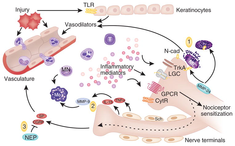
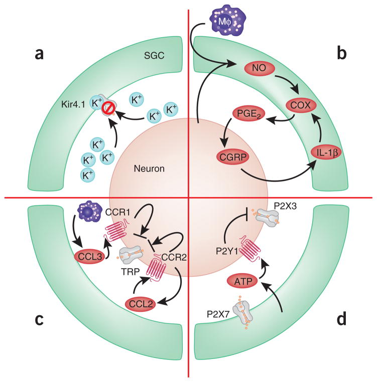
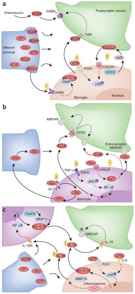
Immune cells and glia interact with neurons to alter pain sensitivity and to mediate the transition from acute to chronic pain. In response to injury, resident immune cells are activated and blood-borne immune cells are recruited to the site of injury. Immune cells not only contribute to immune protection but also initiate the sensitization of peripheral nociceptors. Through the synthesis and release of inflammatory mediators and interactions with neurotransmitters and their receptors, the immune cells, glia and neurons form an integrated network that coordinates immune responses and modulates the excitability of pain pathways. The immune system also reduces sensitization by producing immune-derived analgesic and anti-inflammatory or proresolution agents. A greater understanding of the role of the immune system in pain processing and modulation reveals potential targets for analgesic drug development and new therapeutic opportunities for managing chronic pain.


Conflict of interest statement

Competing Financial Interests

The authors declare no competing financial interests.

Figures

Figure 1
Figure 1
Immune activation and nociceptor sensitization after injury. Injury initiates the release of mediators that activate TLRs on keratinocytes (top) and mast cells (MC) close to the nerve terminal. Vasodilators are also released, promoting adhesion and transmigration of immune cells including T cells (T), neutrophils (N) and monocytes (MN), and recruitment of macrophages (Mφ). These cells, once activated, release a battery of inflammatory mediators that act on receptors expressed on adjacent nociceptor nerve terminals, leading to peripheral nociceptor sensitization. Targets include cytokine receptors (CytR), G protein–coupled receptors (GPCR), ligand-gated channels (LGC) and tyrosine kinase receptor type 1 (TrkA). Three examples of interactions between immune cells and nerve terminals are depicted. (1) Mast cell degranulation requires direct contact between mast cells and nerve terminals, mediated by N-cadherin (N-cad). The metalloproteinase MMP-24 prevents mast cell degranulation by digesting N-cad. (2) Release of TNF-α and IL-15 by peripheral nerves and Schwann cells activates MMP-9 and facilitates recruitment of macrophages. (3) Nociceptive nerve terminals can secrete substance P (SP) and CGRP through antidromic activation of neighboring nerve terminal branches (see text). Substance P and CGRP promote vasodilation and extravasation of immune cells. Neutral endopeptidase (NEP) restrains neuroinflammation by degrading substance P and CGRP.
Figure 2
Figure 2
Modulation of sensory nerve activity in dorsal root ganglia by SGCs. (a) Nerve injury reduces Kir4.1 expression in SGCs, resulting in reduced K+ buffering and increased neuronal excitability. (b) A reciprocal paracrine signaling loop involving NO, COX, PGE2, CGRP and IL-1β. Macrophages infiltrate into the space between SGCs and neurons and secrete inflammatory mediators. (c) Chemokine-mediated regulation of neuronal TRP channels through paracrine (Schwann cell–derived CCL3 and neuronal CCR1) and autocrine (neuron-derived CCL2 and neuronal CCR2) signaling. (d) P2X7R in SGC tonically inhibits P2X3R in neurons by activating neuronal P2Y1.
Figure 3
Figure 3
Activation of glia and neurons in the dorsal horn of the spinal cord after peripheral injury. (a) Microglia-neuron interactions. Upon activation, afferent nerve terminals release neurotransmitters, substance P, CGRP, glutamate (Glu), ATP and BDNF, as well as inflammatory mediators including IL-6 and CCL2 and the growth and differentiation factor neuregulin-1 (NRG-1), into the spinal cord. Three examples are shown. (1) Neuronal NRG-1 acts on microglial erbB2, leading to IL-1β release. (2) Microglial cathepsin S (catS) cleaves neuronal CX3CL1, which binds CX3CR1 and stimulates phosphorylation of p38 MAPK in microglia. This pathway may be inhibited by protein-coupled receptor kinase 2 (GRK2). (3) ATP binds P2X4 and induces BDNF release from microglia, which upon binding TrkB receptor induces a shift in the chloride anion gradient and GABAA receptor-mediated depolarization in dorsal horn neurons. (b) Astrocyte-neuron interactions. (1) Astrocytes release glutamate and D-serine, which bind extrasynaptic and synaptic NMDA receptors on neurons, respectively. (2) Injury-induced downregulation of astrocytic GLT-1 alters glutamate homeostasis in the synaptic cleft. (3) TNF-α activates the JNK1 pathway, which leads to release of CCL2 and alterations in NMDAR and AMPAR activity. (c) Cross-talk between nerve terminals, astrocytes and glia. (1) TLR priming and purinergic signaling increase IL-1β release by glia, which modulates NMDA receptor activity on postsynaptic neurons. TIMPs in astrocytes inhibit MMP-mediated cleavage of pro–IL-1β. (2) Microglial IL-18 binds IL18R on astrocytes and induces NF-κB activity and upregulation of inflammatory cytokines. Dashed lines represent multiple intermediate signaling events.

References

    1. DeLeo JA, Tanga FY, Tawfik VL. Neuroimmune activation and neuroinflammation in chronic pain and opioid tolerance/hyperalgesia. Neuroscientist. 2004;10:40–52. - PubMed
    1. Watkins LR, Maier SF. Immune regulation of central nervous system functions: from sickness responses to pathological pain. J Intern Med. 2005;257:139–155. - PubMed
    1. Scholz J, Woolf CJ. The neuropathic pain triad: neurons, immune cells and glia. Nat Neurosci. 2007;10:1361–1368. - PubMed
    1. Ren K, Dubner R. Neuron-glia crosstalk gets serious: role in pain hypersensitivity. Curr Opin Anaesthesiol. 2008;21:570–579. - PMC - PubMed
    1. Thacker MA, Clark AK, Marchand F, McMahon SB. Pathophysiology of peripheral neuropathic pain: immune cells and molecules. Anesth Analg. 2007;105:838–847. - PubMed

Publication types