. 2010 Dec;11(12):969-76.
doi: 10.1038/embor.2010.154. Epub 2010 Oct 15.

Traf7, a MyoD1 transcriptional target, regulates nuclear factor-κB activity during myogenesis

Affiliations

Traf7, a MyoD1 transcriptional target, regulates nuclear factor-κB activity during myogenesis

Mary Tsikitis et al. EMBO Rep. 2010 Dec.

Abstract

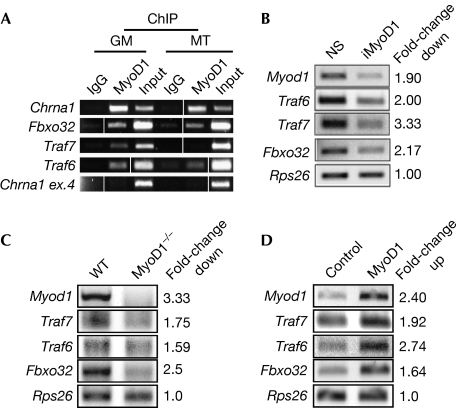
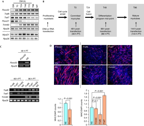
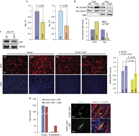
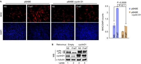
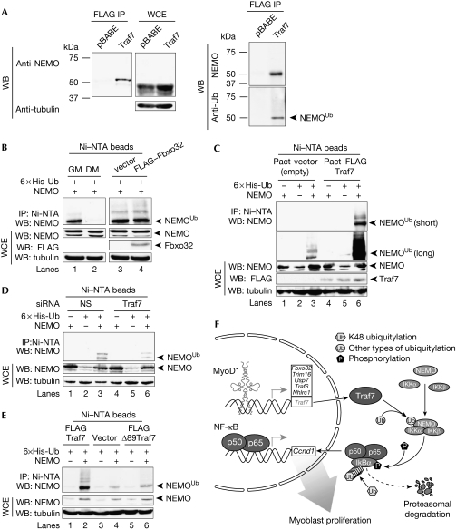
We have identified the E3 ligase Traf7 as a direct MyoD1 target and show that cell cycle exit-an early event in muscle differentiation-is linked to decreased Traf7 expression. Depletion of Traf7 accelerates myogenesis, in part through downregulation of nuclear factor-κB (NF-κB) activity. We used a proteomic screen to identify NEMO, the NF-κB essential modulator, as a Traf7-interacting protein. Finally, we show that ubiquitylation of NF-κB essential modulator is regulated exclusively by Traf7 activity in myoblasts. Our results suggest a new mechanism by which MyoD1 function is coupled to NF-κB activity through Traf7, regulating the balance between cell cycle progression and differentiation during myogenesis.


Conflict of interest statement

The authors declare that they have no conflict of interest.

Figures

Figure 1
Figure 1
MyoD1 regulates ubiquitin-related genes. (A) ChIP validation for MyoD1 binding to selected targets in C2C12 growing myoblasts (GM) and myotubes (MT). Input is 0.5% of total starting material. Chrna1, positive control; Chrna1 exon 4, negative control. Vertical lines indicate that intervening lanes were removed. (B) RT–PCR analysis after MyoD1 RNAi (iMyoD1). NS, nonspecific control. (C) RT–PCR analysis in wild-type (WT) and MyoD1−/− primary myoblasts. (D) RT–PCR analysis after MyoD1 overexpression in growing C2C12 cells. Rps26 is a nonspecific input and loading control. Fold-change in mRNA levels (BD) determined by densitometric quantification using Image J software. ChIP, chromatin immunoprecipitation; mRNA, messenger RNA; NS, nonspecific; RNAi, RNA interference; RT–PCR, reverse transcriptase PCR; WT, wild type.
Figure 2
Figure 2
A distinct function for Traf7 in differentiation. (A) RT–PCR analysis of indicated E3 ligases (top) and MyoD1 (bottom). Rps26, input and loading control. GM and MT indicate growing myoblasts and myotubes, respectively. Cells are confluent at T0. T24–T96 indicate time (hours) in DM. (B) Diagram of experimental scheme. Transfections were performed in growing myoblasts at <50–60% confluence. (C) RT–PCR analysis of Fbxo32 (T0) and Traf6, Traf7 and Trim63 depletion at T0 (48 h PT) or T48 (96 h post-transfection). (D) Top panels show immunofluorescence on myoblasts depleted of Fbxo32 (left) or Traf6, Traf7 or Trim63 (right, independent experiment) and differentiated for 2 days. Scale bar, 40 μm, magnification × 10. Trim63, control; RNAi performed with pool of four siRNAs. Cells were stained with MHC antibodies (red) and DAPI (blue). Quantitation of data in top panel is also shown; MHC/DAP1 signal for each siRNA was normalized against NS. Error bars indicate s.e.m. values; P-values shown for n=5 independent experiments. DAPI, 4′,6-diamidino-2-phenylindole; DM, differentiation medium; MHC, myosin heavy chain; NS, nonspecific; PT, post-transfection; RNAi, RNA interference; RT–PCR, reverse transcriptase PCR.
Figure 3
Figure 3
A pathway connecting Traf7 and p65. (A) NF-κB luciferase reporter assay in growing myoblasts depleted of Traf7 (left) or MyoD1 (right). Luciferase counts were normalized against Renilla to show relative fold changes (FC). (B) Top panel: Immunoblot (IB) analysis of p65 in cytoplasmic (Cyto) and nuclear (Nuc) fractions from lysates of confluent myoblasts. TATA-binding protein (TBP) and α-tubulin represent loading controls for nuclear and cytoplasmic extracts, respectively. Bottom panel: normalized densitometric quantification of top panel. (C) Top panel: RT–PCR analysis of p65 mRNA in myoblasts 48 h after siRNA transfection. Bottom, left: IF of MHC expression (red) in myoblasts transfected with vector or FLAG–Traf7 and depleted of p65 (ip65) then differentiated for 48 h (scale bar, 40 μm; magnification × 10). Bottom, right: Normalized MHC expression for ip65-treated cells compared with NS controls. (D) Quantitation of MHC- and FLAG/MHC-positive cells in the ip65 rescue experiment. Panels show IF of FLAG–Traf7 (green) and MHC (red) in myoblasts transfected with or without p65 siRNA and FLAG–Traf7 and differentiated for 48 h. Scale bars, 5 μm for magnification × 63 and 4.92 μm for magnification × 64. Error bars indicate s.e.m. values; P-values shown for three independent experiments. DAPI, 4′,6-diamidino-2-pheylindole; FC, fold-change; IB, immunoblot; IF, immunofluorescence; MHC, myosin heavy chain; mRNA, messenger RNA; NF, nuclear factor; NS, nonspecific; TBP, TATA-binding protein.
Figure 4
Figure 4
Traf7 expression positively correlates with pRB phosphorylation state. (A) Immunofluorescence of MHC expression in myoblasts ectopically expressing cyclin D1 and depleted of Traf7 (iTraf7) and differentiated for 48 h. Scale bars, 40 μm; magnification × 10. Histogram shows normalized MHC signal in iTraf7 cultures compared with that of control. Error bars indicate s.e.m. values; P-values for three independent experiments. (B) pRB and cyclin D1 immunoblots on 100 μg of lysate. DAPI, 4′,6-diamidino-2-phenylindole; MHC, myosin heavy chain; NS, nonspecific; pRb, hypo-phosphorylated pRb; ppRb, hyper-phosphorylated pRb.
Figure 5
Figure 5
Traf7 specifically enhances ubiquitylation of NEMO in myoblasts. (A) IB analysis of lysates from confluent stable FLAG–Traf7 C2C12 cells. (Left) IB of FLAG IP and whole-cell extract (WCE) probed with NEMO (top; from MBL) and tubulin (bottom) antibodies. (Right) IB was probed with NEMO (top; from NCI) and ubiquitin antibodies (bottom). (B) Lanes 1 and 2, IB of lysates from confluent myoblasts (GM) or myotubes differentiated for 48 h (DM) transiently transfected with NEMO and His6–Ub expression constructs. Lanes 3 and 4, IB of lysates from GM transiently transfected with FLAG–Fbxo32 or vector alone along with NEMO and His6–Ub expression constructs. The latter were included in transfections in (CE). Purification of lysates using Ni–NTA beads under denaturing conditions and input WCE are indicated. (Top) IB with NEMO antibodies. (Bottom) IB of WCE with NEMO, FLAG, and α-tubulin (loading control) antibodies. (C) IB of lysates from GM transfected with FLAG–Traf7 or vector alone. (Top) IB with NEMO antibodies. Two exposures shown. (Bottom) IB as in (C). (D) IB of lysates from GM transfected with Traf7 or control (NS) siRNAs. IB as in (C). Unmodified and modified NEMO are indicated. (E) IB of lysates from GM transiently transfected with vector alone, Traf7, or a Traf7 mutant (Δ89 Traf7), together with NEMO and His6–Ub expression constructs. IB as in (C). (F) Model of process whereby Traf7, a MyoDI transcriptional target, enhances NEMO ubiquitylation in myoblasts. DM, differentiation medium; IB, immunoblot; IP, immunoprecipitation; MBL, MBL International; NCI, National Cancer Institute; NEMO, nuclear factor-κB essential modulator; NS, nonspecific; NTA, nitrilotriacetic acid; siRNA, small interfering RNA; Ub, ubiquitin; WB, western blot.

References

    1. Bakkar N, Wang J, Ladner KJ, Wang H, Dahlman JM, Carathers M, Acharyya S, Rudnicki MA, Hollenbach AD, Guttridge DC (2008) IKK/NF-κB regulates skeletal myogenesis via a signaling switch to inhibit differentiation and promote mitochondrial biogenesis. J Cell Biol 180: 787–802 - PMC - PubMed
    1. Blais A, Tsikitis M, Acosta-Alvear D, Sharan R, Kluger Y, Dynlacht BD (2005) An initial blueprint for myogenic differentiation. Genes Dev 19: 553–569 - PMC - PubMed
    1. Guttridge DC, Albanese C, Reuther JY, Pestell RG, Baldwin AS (1999) NF-κB controls cell growth and differentiation through transcriptional regulation of cyclin D1. Mol Cell Biol 19: 5785–5799 - PMC - PubMed
    1. Ha H, Han D, Choi Y (2009) Traf-mediated TNFR-family signaling. Curr Protoc Immunol Ch. 11, Unit 11.9D - PubMed
    1. Halevy O, Novitch BG, Spicer DB, Skapek SX, Rhee J, Hannon GJ, Beach D, Lassar AB (1995) Correlation of terminal cell cycle arrest of skeletal muscle with induction of p21 by MyoD. Science 267: 1018–1021 - PubMed

Publication types

MeSH terms