. 2010:2010:131630.
doi: 10.4061/2010/131630. Epub 2009 Sep 30.

Shrinkage Stresses Generated during Resin-Composite Applications: A Review

Affiliations

Shrinkage Stresses Generated during Resin-Composite Applications: A Review

Luis Felipe J Schneider et al. J Dent Biomech. 2010.

Abstract

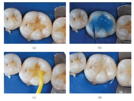
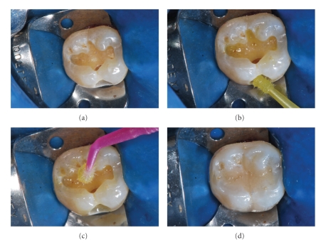
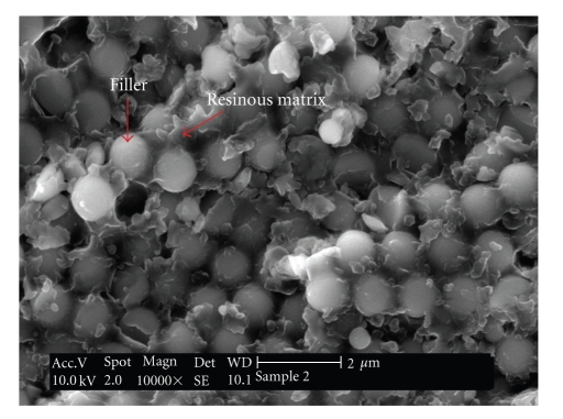
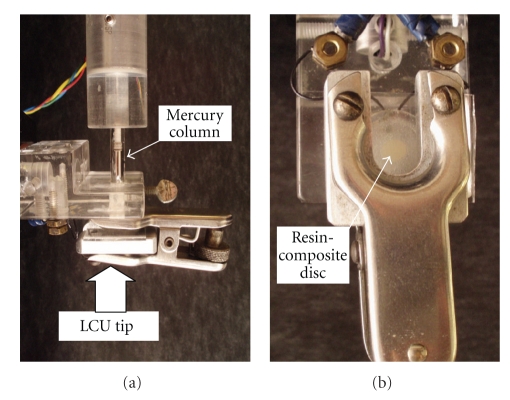
Many developments have been made in the field of resin composites for dental applications. However, the manifestation of shrinkage due to the polymerization process continues to be a major problem. The material's shrinkage, associated with dynamic development of elastic modulus, creates stresses within the material and its interface with the tooth structure. As a consequence, marginal failure and subsequent secondary caries, marginal staining, restoration displacement, tooth fracture, and/or post-operative sensitivity are clinical drawbacks of resin-composite applications. The aim of the current paper is to present an overview about the shrinkage stresses created during resin-composite applications, consequences, and advances. The paper is based on results of many researches that are available in the literature.


Figures

Figure 1
Figure 1
(a) After caries removal, the cavity is prepared to receive the resin-composite restoration. (b) Acid etching treatment with phosphoric acid 37%. (c) Application of a simplified (1 step) adhesive system. (d) After the photoactivation procedure, the resin-composite restoration was built. Adhesive system used: Single-Bond (3MESPE). Resin composite: XRV Ultra (Kerr).
Figure 2
Figure 2
(a) Cavity prepared to receive the resin-composite restoration. (b) Self-etching primer application. (c) Bonding agent application. (d) After the photoactivation procedure, the resin-composite restoration was built. Adhesive system used: Self-etching Silorane (3MESPE). Resin composite: Filtek Silorane (3MESPE).
Figure 3
Figure 3
Scanning electron microscopy (SEM) image of an experimental dental resin composite. It can be easily observed the presence of spherical-shape fillers surrounded by the resin matrix.
Figure 4
Figure 4
Resin monomers often used in the formulations of dental resin composites.
Figure 5
Figure 5
(a) Mercury dilatometer. It can be observed the mercury column, the clasp that holds the resin composite sample (b), and the place where the LCU is positioned. These pictures were kindly donated by Dr. Carmen Silvia C. Pfeifer. Equipment is from the Division of Biomaterials and Biomechanics, School of Dentistry, Oregon Health & Sciences University (Portland, USA).
Figure 6
Figure 6
(a) The “Bonded-disc” apparatus. (b) A close view of the LVDT probe in contact with the glass slide during the resin-composite photoactivation. Equipment is from the Biomaterials Research Group, School of Dentistry, University of Manchester (Manchester, UK).
Figure 7
Figure 7
Extensometer apparatus that is connected to a universal testing machine. As a feedback response, the system compensates deformations and the sample remains constant. Consequently, this kind of method is known as a “low-compliant method.” Pictures kindly donated by Dr. Carmen Silvia C. Pfeifer. Equipment is from the School of Dentistry, University of São Paulo (São Paulo, Brazil).
Figure 8
Figure 8
Controlled compliance apparatus for contraction stress test. (a) The entire apparatus with a view of the steel frame and the upper load cell holder; (b) slot for light guide; (c) glass plate positioned; (d) steel piston in position and the space where the resin-composite specimen is positioned; (e) equipment ready for use; (f) light curing procedure during the experiment. These pictures were kindly donated by Dr. Carmen Silvia C. Pfeifer. Equipment is from Division of Biomaterials and Biomechanics, the School of Dentistry, Oregon Health & Sciences University (Portland, USA).
Figure 9
Figure 9
(a) The Bioman stress measurement device. (b) A close view of the resin-composite specimen. Equipment is from the Biomaterials Research Group, School of Dentistry, University of Manchester (Manchester, UK).

References

    1. Bowen RL. Properties of a silica-reinforced polymer for dental restorations. The Journal of the American Dental Association. 1963;66:57–64. - PubMed
    1. Bowen RL. Adhesive bonding of various materials to hard tooth tissues—VI: forces developing in direct-filling materials during hardening. The Journal of the American Dental Association. 1967;74(3):439–445. - PubMed
    1. Silikas N, Eliades G, Watts DC. Light intensity effects on resin-composite degree of conversion and shrinkage strain. Dental Materials. 2000;16(4):292–296. - PubMed
    1. Causton BE, Miller B, Sefton J. The deformation of cusps by bonded posterior composite restorations: an in vitro study. British Dental Journal. 1985;159(12):397–400. - PubMed
    1. McCullock AJ, Smith BG. In vitro studies of cuspal movement produced by adhesive restorative materials. British Dental Journal. 1986;161(11):405–409. - PubMed

LinkOut - more resources