. 2010 Oct 7;5(10):e13202.
doi: 10.1371/journal.pone.0013202.

Structural transformation of the tandem ubiquitin-interacting motifs in ataxin-3 and their cooperative interactions with ubiquitin chains

Affiliations

Structural transformation of the tandem ubiquitin-interacting motifs in ataxin-3 and their cooperative interactions with ubiquitin chains

Ai-Xin Song et al. PLoS One. .

Abstract

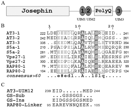
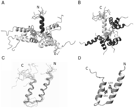
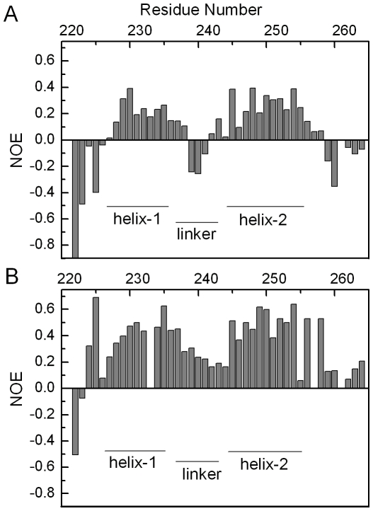
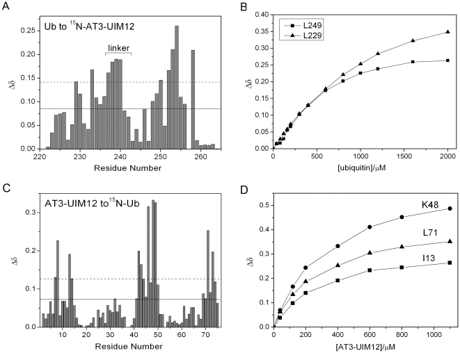
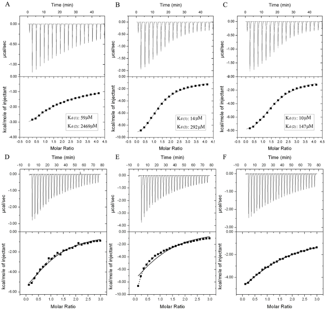
The ubiquitin-interacting motif (UIM) is a short peptide with dual function of binding ubiquitin (Ub) and promoting ubiquitination. We elucidated the structures and dynamics of the tandem UIMs of ataxin-3 (AT3-UIM12) both in free and Ub-bound forms. The solution structure of free AT3-UIM12 consists of two α-helices and a flexible linker, whereas that of the Ub-bound form is much more compact with hydrophobic contacts between the two helices. NMR dynamics indicates that the flexible linker becomes rigid when AT3-UIM12 binds with Ub. Isothermal titration calorimetry and NMR titration demonstrate that AT3-UIM12 binds diUb with two distinct affinities, and the linker plays a critical role in association of the two helices in diUb binding. These results provide an implication that the tandem UIM12 interacts with Ub or diUb in a cooperative manner through an allosteric effect and dynamics change of the linker region, which might be related to its recognitions with various Ub chains and ubiquitinated substrates.


Conflict of interest statement

Competing Interests: The authors have declared that no competing interests exist.

Figures

Figure 1
Figure 1. Domain Architecture of AT3 Protein and Sequence Alignments of the UIM Motifs.
A, Domain architecture of AT3 protein. B, Multiple sequence alignments of the UIM motifs from AT3, S5a, Vps27 and RAP80. Note that the length of the linker region between the two tandem UIM motifs varies in these four proteins. The linker in AT3 is the shortest (6 residues), and then in RAP80 (∼11 residues), whereas those in S5a and Vps27p are longer with 50 and 23 residues, respectively. The sequences were aligned using ClustalW and represented using ESPript. C, Sequences of the linker regions for wild-type AT3-UIM12 and its mutants. GS-Sub, substitution with GSSGGS sequence; GS-Ins, Insertion of GSSGGS; RAP80-Linker, replacement with the linker sequence of RAP80.
Figure 2
Figure 2. Solution Structures of Tandem AT3-UIM12 in Free and Ub-Bound Forms.
A, Ensemble of 10 structures superimposed on the polypeptide backbones of the α-helix of UIM1. B, Ensemble of 10 structures superimposed on those of UIM2. The helical portions of AT3-UIM12 show relative high definition, but the linker region is unstructured and flexible. No tertiary contacts between helices are observed. C, Backbone superposition of 10 lowest-energy structures of AT3-UIM12 in the Ub-bound form. D, Ribbon representation of AT3-UIM12 structure in the Ub-bound form. The structure of the Ub-bound form includes two α-helices, helix-1 (residues 226–238) and helix-2 (residues 247–256), and a linker loop region (residues 239–244) between them. The figures were generated using MOLMOL.
Figure 3
Figure 3. Backbone Dynamics of AT3-UIM12 in the Free and Ub-Bound Forms.
A, The 15N-{1H}-NOE values of backbone amides are plotted against the residue number of AT3-UIM12 in the absence Ub. B, The 15N-{1H}-NOE values in the presence of two-fold amount of Ub. The secondary structures of AT3-UIM12 are indicated as narrow bars.
Figure 4
Figure 4. Interaction of Tandem AT3-UIM12 with Ub.
A, Diagram of the chemical shift changes (Δδ) of AT3-UIM12 against residue number at an AT3-UIM12/Ub molar ratio of 1∶2. B, Titration curves for two equivalent residues (Leu229 in UIM1 and Leu249 in UIM2) in tandem AT3-UIM12 upon Ub binding. Note that the Δδ value of Leu249 in UIM2 upon Ub titration reaches the plateau with lower amount of Ub than the corresponding Leu229 in UIM1. C, Diagram of the chemical shift changes (Δδ) of Ub against residue number at an Ub/AT3-UIM12 molar ratio of 1∶2. D, Titration curves for three residues (Ile13, Lys48 and Leu71) in Ub upon GB1-tagged AT3-UIM12 binding. The dissociation constant for the binding of AT3-UIM12 with Ub is listed in Table 1. The solid and dashed lines (A & C) indicate the threshold values of mean and mean plus SD for the chemical shift changes.
Figure 5
Figure 5. AT3-UIM12 Binds with Ub Through Hydrophobic Interactions.
A, Mapping of the significantly perturbed residues by AT3-UIM12 interaction on Ub surface. The residues with chemical shift changes larger than the average have been labeled in pink and those larger than the average plus SD in red (ref. Fig. 3C). The Ub structure is referenced from the crystal structure (PDB code: 1UBQ). B, Intermolecular NOEs observed in the AT3-UIM12-Ub complex. A 1∶2 molar ratio of labeled AT3-UIM12 to unlabeled Ub were mixed for 3D 13C-filtered/15N-edited NOESY spectrum. The figure shows the NOE peaks projected onto the 2D plane. C, Mapping of the residues that have the intermolecular NOEs with Ub on the ribbon diagram of AT3-UIM12. D, Mapping of the residues that have the intermolecular NOEs with Ub on the surface diagram of AT3-UIM12, showing two patches for binding with two Ub molecules. The ribbon and surface representations were generated by MOLMOL.
Figure 6
Figure 6. The Cooperative Interaction of AT3-UIM12 with K48- and K63- Linked diUb Chains.
A, Pull-down of K48- and K63-linked diUb by GST-fused UIM1, UIM2 or UIM12. The pull-down samples were detected by Western blotting with an anti-Ub antibody. B, SDS-PAGE of the GST-fused proteins (AT3-UIM1, AT3-UIM2 and AT3-UIM12) detected by Coomassie staining.
Figure 7
Figure 7. Isothermal Titration Calorimetry Experiments for the Interactions of various AT3-UIM12 forms with diUb types.
A, B, C, Titration of different types of diUb with GB1-AT3-UIM12. A, Linear diUb; B, K48-linked diUb; C, K63-linked diUb. By fitting the data with the sequential binding model, these titrations are well-fitted to give two distinct binding affinities. D, E, F, Titration of K48-linked diUb with different AT3-UIM12 forms. D, GS-Sub; E, GS-Ins; F, RAP80-Linker. The concentration of K48-linked diUb is 100 µM and those of the AT3-UIM12 stocks are 2 mM. The titrations are well-fitted to a usual two-site binding model (dashed line) rather than the sequential binding model (solid line). The concentrations of diUb types are 100 µM and those of AT3-UIM12 stocks are 2 mM. The GB1 fusion was used for peptide expression and quantification.
Figure 8
Figure 8. Schematic Representation of the Tandem UIM Motifs with Various Forms of Ub Chains.
A, Non-cooperative binding mode. A representative for the mode is the tandem UIM motifs in Vps27p , which bind with Ub independently and non-cooperatively. B, Avid binding mode. The tandem UIM motifs in RAP80 bind with K63-linked diUb by forming a long α-helical structure . The predicted linker becomes rather rigid and may be preferable to the configuration of K63-linked diUb. C, Cooperative binding mode. A short linker region (such as in AT3-UIM12) tends to be involved in the cooperative binding with Ub chains or ubiquitinated substrates. This cooperation is also likely to be occurred in poly- or multiUb. When the tandem UIM motifs bind with any form of Ub, a conformational arrangement and dynamics change occur in the linker region that may favor Ub binding. The dashed line indicates that either mono- or diUb is involved in the binding mode, and Ubi denotes different forms of Ub, standing for mono-, di-, poly- or multiUb.

References

    1. Hofmann K, Falquet L. A ubiquitin-interacting motif conserved in components of the proteasomal and lysosomal protein degradation systems. Trends Biochem Sci. 2001;26:347–350. - PubMed
    1. Miller SL, Malotky E, O'Bryan JP. Analysis of the role of ubiquitin-interacting motifs in ubiquitin binding and ubiquitylation. J Biol Chem. 2004;279:33528–33537. - PubMed
    1. Young P, Deveraux Q, Beal RE, Pickart CM, Rechsteiner M. Characterization of two polyubiquitin binding sites in the 26 S protease subunit 5a. J Biol Chem. 1998;273:5461–5467. - PubMed
    1. Oldham CE, Mohney RP, Miller SL, Hanes RN, O'Bryan JP. The ubiquitin-interacting motifs target the endocytic adaptor protein epsin for ubiquitination. Curr Biol. 2002;12:1112–1116. - PubMed
    1. Polo S, Sigismund S, Faretta M, Guidi M, Capua MR, et al. A single motif responsible for ubiquitin recognition and monoubiquitination in endocytic proteins. Nature. 2002;416:451–455. - PubMed

Publication types

MeSH terms