. 2010 Oct 7;6(10):e1001141.
doi: 10.1371/journal.ppat.1001141.

Parvovirus minute virus of mice induces a DNA damage response that facilitates viral replication

Affiliations

Parvovirus minute virus of mice induces a DNA damage response that facilitates viral replication

Richard O Adeyemi et al. PLoS Pathog. .

Abstract

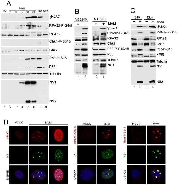
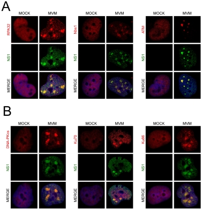
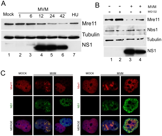
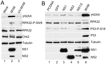
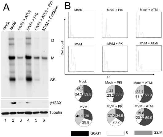
Infection by DNA viruses can elicit DNA damage responses (DDRs) in host cells. In some cases the DDR presents a block to viral replication that must be overcome, and in other cases the infecting agent exploits the DDR to facilitate replication. We find that low multiplicity infection with the autonomous parvovirus minute virus of mice (MVM) results in the activation of a DDR, characterized by the phosphorylation of H2AX, Nbs1, RPA32, Chk2 and p53. These proteins are recruited to MVM replication centers, where they co-localize with the main viral replication protein, NS1. The response is seen in both human and murine cell lines following infection with either the MVMp or MVMi strains. Replication of the virus is required for DNA damage signaling. Damage response proteins, including the ATM kinase, accumulate in viral-induced replication centers. Using mutant cell lines and specific kinase inhibitors, we show that ATM is the main transducer of the signaling events in the normal murine host. ATM inhibitors restrict MVM replication and ameliorate virus-induced cell cycle arrest, suggesting that DNA damage signaling facilitates virus replication, perhaps in part by promoting cell cycle arrest. Thus it appears that MVM exploits the cellular DNA damage response machinery early in infection to enhance its replication in host cells.


Conflict of interest statement

The authors have declared that no competing interests exist.

Figures

Figure 1
Figure 1. MVM infection induces a DNA damage response (DDR).
(A) Time course of DDR following infection of A9 cells. A9 cells were para-synchronized in G0 as described in materials and methods. Cells were then mock-infected or infected with MVMp at an MOI of 7. As a positive control for the DDR, A9 cells were treated with 2 mM hydroxyurea (HU) for 12 hrs. Infected cells were harvested every 6 hrs over a 24 hr period and lysed in modified RIPA buffer. Protein content was measured using Bradford assay and equal amounts of protein were loaded in each well for immunoblotting. Western blot analysis was carried out using antibodies against NS1/2, tubulin, γH2AX, RPA32, Chk2, phosphor-Chk1, phosphor-p53 and p53. M0 and M24 represents mock samples at 0 hr and 24 hr time points respectively. (B) DDR following MVM infection in murine NIH3T6 cells and permissive human NB324K cells. Western blot analysis of virus and DDR proteins 24 h.p.i. of NIH3T6 cells and NB324K cells with MVMp. For detection of NS1, an antibody specific to NS1 alone was used. (C) DDR following infection with lymphocytic strain of MVM, MVMi. Western blot analysis of DDR proteins 24 h.p.i. of mouse S49 and EL4 lymphocyte cell lines with MVMi. (D) Cellular DDR proteins localized within APAR bodies. Para-synchronized A9 cells were infected with MVMp (MOI of 10) for 24 hrs before being fixed and processed for immunofluorescence. Cells were stained with the indicated antibodies to mark activated DDR proteins. Staining with an antibody to phosphorylated H2AX (γH2AX) was observed in distinct foci and also in a pan-nuclear pattern as described in the text. APAR bodies were detected with antibodies to NS1. Nuclei were stained with DAPI. All images were captured using an objective of 63×.
Figure 2
Figure 2. DNA repair proteins accumulate at MVM APAR bodies.
(A) Repair proteins accumulate at APAR bodies. NB324K cells were infected with MVMp (MOI of 10) for 16 hr before being fixed and processed for immunofluorescence. Cells were stained with the indicated antibodies to mark DDR repair proteins. APAR bodies were detected with antibodies to NS1. Nuclei were stained with DAPI. All images were captured using an objective of 63×. (B) DNA-PK components localize to APAR bodies. NB324K cells were infected with MVMp (MOI of 10) for 24 hr before being fixed and processed for immunofluorescence. Cells were stained with the indicated antibodies. APAR bodies were detected with antibodies to NS1. Nuclei were stained using DAPI. All images were captured using an objective of 63×.
Figure 3
Figure 3. Mre11 but not Nbs1 is degraded in MVM-infected cells.
(A) Mre11 levels were reduced late in infection. Western blot analysis of a time course of MVM infection of A9 cells shows reduction in the levels of Mre11 protein at 24 h.p.i but not in mock-infected (M) or hydroxyurea-treated (HU) cells. Tubulin served as a loading control. (B) Mre11 loss is proteasome-dependent. Mock-infected and MVM-infected A9 cells were treated with MG132 or DMSO at 24 h.p.i. for 6 hours. Western blots show reversal of Mre11 loss following treatment with MG132, whereas the levels of Nbs1 remain unchanged. (C) Mre11 loss examined by immunofluorescence. Confocal immunofluorescent microscopy comparing staining for Mre11 and Nbs1 in uninfected and MVM-infected NB324K cells. Both Mre11 and Nbs1 are found co-localized with NS1 in APAR bodies at early stages of infection but only Mre11 levels are specifically decreased at late stages of infection when NS1 is diffusely nuclear. All images were captured using an objective of 63×.
Figure 4
Figure 4. MVM-induced DDR requires viral replication.
(A) Loss of DDR following UV-inactivation of MVM. MVM was UV-inactivated as described in materials and methods. A9 cells were mock-infected or infected with wild-type or UV-inactivated MVM at an MOI of 10. Western blot analysis of DDR proteins as well as NS1/2 and tubulin, of samples taken 24 hrs post-infection, revealed that UV-inactivated MVM did not activate the signaling pathways induced by wild-type MVM. (B) Expression of non-structural proteins is not sufficient for full DDR. Western blot analysis of DDR proteins 48 hrs following transfection of A9 cells with plasmids expressing MVM non-structural proteins as well as control plasmids pcDNA and GFP. MVM infection served as a positive control for the DDR, and tubulin was used as a loading control.
Figure 5
Figure 5. Activated ATM is predominantly responsible for signaling events during MVM infection.
(A) ATM inhibitor reduces signaling events. Western blot analysis comparing DDR proteins at 18 h.p.i. of parasynchronized A9 cells infected with MVMp in the absence or presence of 7.5 µM ATM inhibitor KU 55933 (ATMi). (B) ATM-mediated signaling events seen in the absence of DNA-PKcs. Western blot analysis of DDR proteins following mock or MVMp infection of CHO-AA8 and CHO-V3 cells deficient in DNA-PKcs. Treatment with 10 µM ATM inhibitor KU 55933 (ATMi) blocked signaling in the CHO-V3 cells. (C) Activation of ATM following MVM infection. Western blot analysis shows ATM phosphorylation on S1981 following MVM infection of NB324K cells and hydroxyurea (HU) treatment. (D) ATM-dependent staining at APAR bodies. NB324K cells were infected with MVMp (MOI of 10) for 16 hr before being fixed and processed for immunofluorescence. Cells were stained with antibodies to NS1 and an antibody generated to phosphorylated ATM. Nuclei were stained with DAPI. All images were captured using an objective of 63×.
Figure 6
Figure 6. ATM kinase activity is required for efficient MVM replication.
(A) Reduction of virus replication following inhibition of ATM kinase activity. A9 cells were pre-treated with DMSO or 7.5 µM ATM inhibitor (ATMi), 10 µM DNA-PK inhibitor (PKi), ATM and DNA-PK inhibitors in combination (ATMi+PKi), or 2.5 mM caffeine for 30 min prior to MVM infection at an MOI of 10. Lysates were split in half and used for Southern and western blot analysis. Southern blots were carried out as described in materials and methods and shown in the upper panel. DNA content was measured and equal amount of DNA loaded in each well. The blot was hybridized with a radiolabeled MVM probe and replicative intermediates of single-stranded DNA, SS; monomer, M; and dimer, D; are indicated to the right. Western blot analysis was also carried out as described and shown in the lower panel. The reduction in γH2AX signal confirmed inhibition of signaling by the inhibitors. (B) Reversal of cell cycle arrest following ATM inhibition. FACS analysis was carried out as described in materials and methods. Cells were pre-treated for one hour with 7.5 µM KU55933 (ATMi), 10 µM NU7026 (DNA-PKi) or DMSO vehicle before being mock-infected or infected with MVMp at an MOI of 10 for 28 hours. Representative histograms showing cell counts plotted against propidium iodide (PI) intensity are shown. The percentage of cells in each cycle stage was quantified using Flowjo software and shown in pie charts. Numbers are averages from two independent experiments.

References

    1. Lilley CE, Schwartz RA, Weitzman MD. Using or abusing: viruses and the cellular DNA damage response. Trends Microbiol. 2007;15:119–126. - PubMed
    1. Weitzman MD, Carson CT, Schwartz RA, Lilley CE. Interactions of viruses with the cellular DNA repair machinery. DNA Repair (Amst) 2004;3:1165–1173. - PubMed
    1. Evans JD, Hearing P. Relocalization of the Mre11-Rad50-Nbs1 complex by the adenovirus E4 ORF3 protein is required for viral replication. J Virol. 2005;79:6207–6215. - PMC - PubMed
    1. Carson CT, Orazio NI, Lee DV, Suh J, Bekker-Jensen S, et al. Mislocalization of the MRN complex prevents ATR signaling during adenovirus infection. Embo J. 2009;28:652–662. - PMC - PubMed
    1. Lakdawala SS, Schwartz RA, Ferenchak K, Carson CT, McSharry BP, et al. Differential requirements of the C terminus of Nbs1 in suppressing adenovirus DNA replication and promoting concatemer formation. J Virol. 2008;82:8362–8372. - PMC - PubMed

Publication types

MeSH terms