. 2010 Oct 7;6(10):e1001142.
doi: 10.1371/journal.ppat.1001142.

Requirements for receptor engagement during infection by adenovirus complexed with blood coagulation factor X

Affiliations

Requirements for receptor engagement during infection by adenovirus complexed with blood coagulation factor X

Angela C Bradshaw et al. PLoS Pathog. .

Abstract

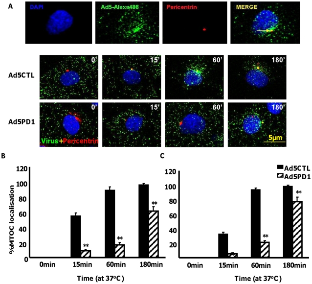
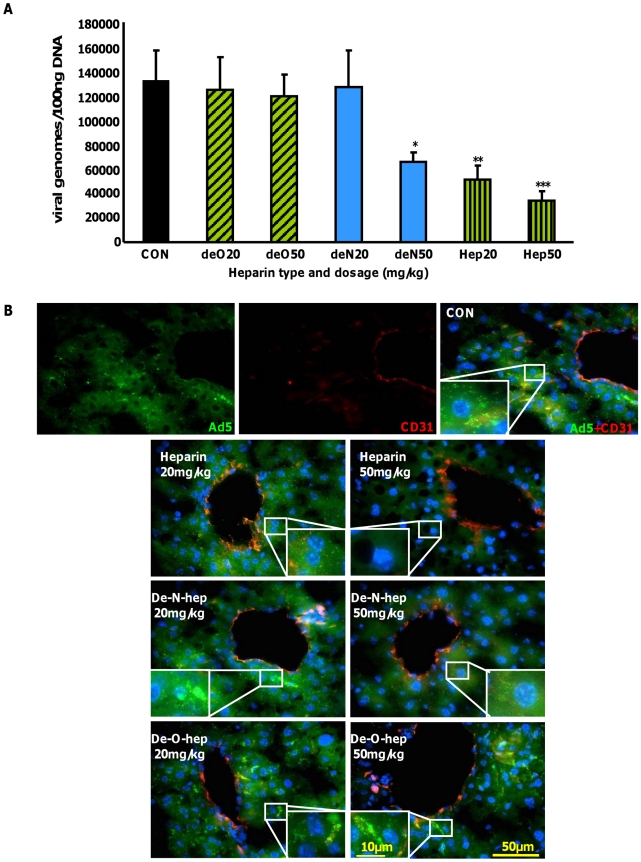
Human adenoviruses from multiple species bind to coagulation factor X (FX), yet the importance of this interaction in adenovirus dissemination is unknown. Upon contact with blood, vectors based on adenovirus serotype 5 (Ad5) binds to FX via the hexon protein with nanomolar affinity, leading to selective uptake of the complex into the liver and spleen. The Ad5:FX complex putatively targets heparan sulfate proteoglycans (HSPGs). The aim of this study was to elucidate the specific requirements for Ad5:FX-mediated cellular uptake in this high-affinity pathway, specifically the HSPG receptor requirements as well as the role of penton base-mediated integrin engagement in subsequent internalisation. Removal of HS sidechains by enzymatic digestion or competition with highly-sulfated heparins/heparan sulfates significantly decreased FX-mediated Ad5 cell binding in vitro and ex vivo. Removal of N-linked and, in particular, O-linked sulfate groups significantly attenuated the inhibitory capabilities of heparin, while the chemical inhibition of endogenous HSPG sulfation dose-dependently reduced FX-mediated Ad5 cellular uptake. Unlike native heparin, modified heparins lacking O- or N-linked sulfate groups were unable to inhibit Ad5 accumulation in the liver 1h after intravascular administration of adenovirus. Similar results were observed in vitro using Ad5 vectors possessing mutations ablating CAR- and/or α(v) integrin binding, demonstrating that attachment of the Ad5:FX complex to the cell surface involves HSPG sulfation. Interestingly, Ad5 vectors ablated for α(v) integrin binding showed markedly delayed cell entry, highlighting the need for an efficient post-attachment internalisation signal for optimal Ad5 uptake and transport following surface binding mediated through FX. This study therefore integrates the established model of α(v) integrin-dependent adenoviral infection with the high-affinity FX-mediated pathway. This has important implications for mechanisms that define organ targeting following contact of human adenoviruses with blood.


Conflict of interest statement

The authors have declared that no competing interests exist.

Figures

Figure 1
Figure 1. Importance of HS sidechains during Ad5 binding, cell entry, cytosolic transport and transduction in vitro.
(A) Binding of 1000 vp/cell Ad5CTL, Ad5KO1(CAR-binding mutant), Ad5PD1 (αv integrin-binding mutant) or Ad5KP(CAR- and αv integrin-binding mutant) particles to cells was quantified after incubation of Ad particles with HepG2 or SKOV3 cells for 1 h at 4°C in the presence or absence of 10 µg/ml FX. Vector genomes were detected by quantitative PCR. (B+C) SKOV3 (left panels) or HepG2 (right panels) cells were incubated for 1 hour at 37°C in serum free media containing 0–5 U/ml of Heparinase III. The effect of heparinase pre-treatment on FX-mediated Ad5 binding (1000 vp/cell, 1 hour, 4°C) (B) or Ad5 mediated transduction (1000 vp/cell, 3 hours, 37°C) was quantified (C). ** = p<0.01. (D) (upper panel) 10,000 vp/cell of Alexa488-labelled Ad5 (green) were allowed to bind cells for 1 h at 4°C, then incubated at 37°C for 60 min prior to fixation and staining for the MTOC marker pericentrin (red). Nuclei were counterstained using DAPI. Colocalisation of fluorescently-labelled Ad5 particles with pericentrin is indicated by the yellow arrows. Images were captured on a confocal microscope under a 63× objective (D) (lower panel) Alexa-labelled Ad5 was allowed to bind cells for 1 h at 4°C in the presence or absence of 10 µg/ml FX and/or heparin (Hep). Cells were then incubated at 37°C for 0 min to 60 min prior to fixation. Nuclei were counterstained using DAPI. (E) Fluorescently-labelled Ad5 was allowed to bind cells for 1 h at 4°C in the presence or absence of 10 µg/ml FX and heparin (Hep). Nuclei were counterstained using DAPI. Images were captured under a 40× objective.
Figure 2
Figure 2. Endosomal localisation of αv integrin binding-defective Ad5 in vitro.
(A+B) 10,000 vp/cell of Alexa488-labelled Ad5CTL or Ad5PD1 (green particles) in the presence of FX were allowed to bind cells for 1 h at 4°C, then incubated at 37°C for increasing lengths of time (15 min, 60 min) to allow internalisation and trafficking prior to fixation and staining for the early endosome markers EEA1 (A) or Rab5 (B). Specific binding of primary antibodies against EEA1 (A, upper panel) or Rab5 (B, upper panel) was detected using a Alexa546-labelled secondary antibody (red). Nuclei were counterstained using DAPI. Images were captured on a confocal microscope under a 63× objective.
Figure 3
Figure 3. Delayed FX-mediated transport of αv integrin binding-defective Ad5 in vitro.
10,000 vp/cell of Alexa488-labelled Ad5CTL or Ad5PD1 (green particles) in the presence of FX were allowed to bind cells for 1 h at 4°C, then incubated at 37°C for increasing lengths of time (15 min, 60 min, 180 min) to allow internalisation and transport prior to fixation and staining for the MTOC marker pericentrin (A). Specific binding of a primary antibody against pericentrin (A, upper panel) was detected using an Alexa546-labelled secondary antibody (red). Nuclei were counterstained using DAPI. Images were captured on a confocal microscope under a 63× objective. (B+C) Percentage of cells with colocalisation of fluorescently-labelled Ad5CTL or Ad5PD1 with the MTOC marker pericentrin in SKOV3 (B) or A549 cells (C) was calculated by analysing at least 5 separate 40× microscope fields per experimental condition. ** = p<0.01 compared to Ad5CTL values, error bars represent S.E.M.
Figure 4
Figure 4. Effect of shRNA depletion of αv integrin on FX mediated Ad5 transport.
Knockdown of αv integrin mRNA and protein expression levels in SKOV3 cells was quantified by qPCR (A) and flow cytometry (B), respectively. The effect of shRNA depletion on Ad5 trafficking in SKOV3 cells was assessed as previously. Representative images are shown (B) and quantification of peri-nuclear Ad5CTL localisation was calculated by analysis of 5 separate microscope fields per experimental condition (C). **p<0.05 vs both control conditions. (D) Percentage of SKOV3 cells with colocalisation of fluorescently-labelled Ad5CTL in the perinuclear region was calculated by analysing at least 5 separate 40× microscope fields per experimental condition. ** = p<0.01 compared to off target shRNA values, error bars represent S.E.M.
Figure 5
Figure 5. Importance of HSPG sulfation for FX-mediated Ad transduction in vitro.
(A) HepG2 or SKOV3 cells that had been pretreated with increasing concentrations of the sulfation inhibitor sodium chlorate were transduced with 1000 vp/cell of Ad5CTL in the presence (closed bars) or absence (open bars) of 10 µg/ml FX. Pretreated cells were incubated with virus for 3 h at 37°C. Reporter gene activity was quantified 48 h post-transduction. * p<0.05, **p<0.01 compared to control values (0 mM). (B+C) Control CHO-K1, HS-deficient CHO-pgsA745, sulfation-low CHO-pgsE606 or 2-O-sulfation-deficient CHO-pgsF17 were transduced with 1000 vp/cell of Ad5CTL or Ad5PD1 in the presence or absence of 10 µg/ml FX. (B) Cell binding of Ad particles was quantified after incubation for 1 h at 4°C. Vector genomes were detected by quantitative PCR as described previously. (C) Ad5CTL or Ad5PD1 was incubated with cells for 3 h at 37°C. Reporter gene activity was quantified 48 hours post-infection. * p<0.05, **p<0.01 compared to -FX values.
Figure 6
Figure 6. The inhibitory effects of heparin on FX-Ad uptake in vitro and ex vivo are sulfation-dependent.
(A) SKOV3 cells were transduced with 1000 vp/cell of Ad5 in the presence or absence of 10 µg/ml FX and varying concentrations of heparins/heparan sulfates for 3 h at 37°C. Reporter gene expression was quantified 48 h post-transduction. IC50 (µg/ml, inset) values were calculated using the Hill-Slope model. *p<0.05, **p<0.01 compared to CON+FX (i.e. no heparin) values. (B) Liver slices from MF1 mice were incubated with 1×10⁸ vp of Alexa488-labelled Ad5CTL (green) in the presence or absence of 10 µg/ml FX and increasing concentrations of heparins for 1 h at 4°C. Nuclei were counterstained using DAPI. Images were captured using a 60× microscope objective. (C) Attachment of Alexa488-labelled Ad5CTL particles to mouse liver slices were quantitated using the automated cell counting function in ImageJ. Data represent the average number of particles per 60× microscope field. #p<0.01 vs FX− conditions, ** = p<0.01 vs FX+ conditions in the absence of heparin.
Figure 7
Figure 7. Effect of modified heparins on Ad5 liver accumulation.
(A) Heparin or modified heparins were administered to macrophage-depleted MF1 mice 5 minutes prior to intravenous injection of 1×10¹¹ vp of Ad5. Mice were sacrificed 1 h later and Ad genomes were detected in liver tissue lysates by quantitative PCR as previously described (n = 7). *p<0.05, **p<0.01. (B) Control (no heparin), heparin or modified heparins were administered to macrophage-depleted MF1 outbred mice 5 minutes prior to intravenous injection of 1×10¹¹ vp of fluorescently-labelled Ad5 (green). Mice were sacrificed 1 h later and livers were fixed then stained for the endothelial marker CD31 to facilitate imaging of the sinusoids in the liver (red). Nuclei were counterstained using DAPI. Images were captured using a 40× microscope objective.

References

    1. Russell WC. Adenoviruses: update on structure and function. J Gen Virol. 2009;90:1–20. - PubMed
    1. Hong JY, Lee HJ, Piedra PA, Choi EH, Park KH, et al. Lower respiratory tract infections due to adenovirus in hospitalized Korean children: epidemiology, clinical features, and prognosis. Clin Infect Dis. 2001;32:1423–1429. - PubMed
    1. Echavarria M. Adenoviruses in immunocompromised hosts. Clin Microbiol Rev. 2008;21:704–715. - PMC - PubMed
    1. Kojaoghlanian T, Flomenberg P, Horwitz MS. The impact of adenovirus infection on the immunocompromised host. Rev Med Virol. 2003;13:155–171. - PubMed
    1. Blackwell Munsgardt. Adenovirus. Am J Transplant. 2004;4(Suppl 10):101–104. - PubMed

Publication types

MeSH terms