Case Reports
. 2010 Nov;152A(11):2775-83.
doi: 10.1002/ajmg.a.33093.

Natural history of Christianson syndrome

Affiliations
Case Reports

Natural history of Christianson syndrome

Richard J Schroer et al. Am J Med Genet A. 2010 Nov.

Abstract

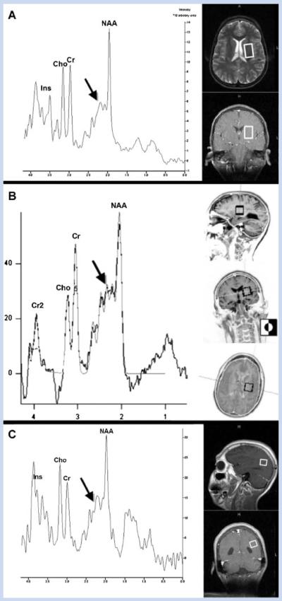
Christianson syndrome is an X-linked mental retardation syndrome characterized by microcephaly, impaired ocular movement, severe global developmental delay, hypotonia which progresses to spasticity, and early onset seizures of variable types. Gilfillan et al.2008] reported mutations in SLC9A6, the gene encoding the sodium/hydrogen exchanger NHE6, in the family first reported and in three others. They also noted the clinical similarities to Angelman syndrome and found cerebellar atrophy on MRI and elevated glutamate/glutamine in the basal ganglia on MRS. Here we report on nonsense mutations in two additional families. The natural history is detailed in childhood and adult life, the similarities to Angelman syndrome confirmed, and the MRI/MRS findings documented in three affected boys.


Figures

FIG. 1
FIG. 1
Partial pedigree of Family 1 (K8310), showing six affected males in two generations and three carrier females in three generations.
FIG. 2
FIG. 2
Facial appearance of five of the six affected males in Family 1 (K8310) and the single affected male in Family 2 (K9524). III-9 at age 35 years showing frontal upsweep, strabismus, open mouth and prominent jaw. III-12 at age 28 years showing microcephaly, upsweep of frontal hair, thin and gaunt face, low nasal septum, open mouth and abnormal hand posturing. IV-1 at age 15 years showing microcephaly and hypotonic face with open mouth. IV-4 at age 611/12 years showing microcephaly and frontal upsweep of hair, but otherwise normal face. IV-6 at age 14 months showing microcephaly, upswept frontal hair, triangular face, wide mouth and thin upper lip. The isolated case in Family 2 is shown at age 6 years with microcephaly and hypotonic face with open mouth.
FIG. 3
FIG. 3
Bilateral abnormal horizontal eye movements in IV-I (family 1, K831) when looking to the right (A) and to the left (B). Although there were occasions that the eyes could almost be laterally buried, the majority of the time he exhibited a bilateral cranial nerve VI paresis on examination.
FIG. 4
FIG. 4
Brain magnetic resonance imaging on maternal half-brothers (IV-1 and IV-4, Family 1, K8310) using a 1.5 T magnet. A,B: Sagittal T1-weighted and coronal FLAIR images respectively on IV-4 at age 7 years. C,D: sagittal T1-weighted and coronal FLAIR images on IV-1 at age 14 years. A,C: Volume loss secondary to focal atrophy and/or hypoplasia of the inferior cerebellar vermis with prominent cerebral spinal fluid space at the cisterna magna in both patients (curved white arrows). The volume loss secondary to focal atrophy and/or hypoplasia is more conspicuous in the oldest patient, IV-1. B,D: Subtle abnormally high signal in the inferior aspect of the cerebellar hemispheres on coronal FLAIR sequences in IV-4 and IV-1 (straight white arrows) which may indicate some degree of gliosis.
FIG. 5
FIG. 5
Transverse spin-echo T2-weighted magnetic resonance images of the brain. A,B: Prominent lateral ventricles in IV-1. Brain MRI coronal FLAIR (Fluid Attenuation Inversion Recover) images. C,D: Mild reduction in size and increase signal of the hippocampi (white arrows image C) in IV-1 in comparison to image D which shows normal size and signal of the hippocampi on IV-4 (black arrows).
FIG. 6
FIG. 6
Single voxel short echo time magnetic resonance spectroscopy (MRS) performed in a 1.5 T magnet (TE: 25–30 ms) demonstrated a strikingly similar abnormally prominent Glutamate/Glutamine shoulder (arrows) in all three spectra. The volumes of interest are located in the left basal ganglia in IV-4 (A), the left basal ganglia in IV-1 (B), and the left parietal white matter in IV-1 (C). The illustrations in the panels on the right indicate the position of the voxels. Standard spectral peaks are illustrated for inositol (Ins), choline (Cho), creatine (Cr), and N-acetylaspartate (NAA).

References

    1. Cavus I, Pan JW, Hetherington HP, Abi-Saab W, Zaveri HP, Vives KP, Krystal JH, Spencer SS, Spencer DD. Decreased hippocampal volume on MRI is associated with increased extracellular glutamate in epilepsy patients. Epilepsia. 2008;49:1358–1366. - PubMed
    1. Christianson AL, Stevenson RE, van der Meyden CH, Pelser J, Theron FW, van Rensburg PL, Chandler M, Schwartz CE. X-linked severe mental retardation, craniofacial dysmorphology, epilepsy, ophthalmoplegia, and cerebellar atrophy in a large South African kindred is localized to Xq24–q27. J Med Genet. 1999;36:759–766. - PMC - PubMed
    1. Gilfillan GD, Selmer KK, Roxrud I, Smith R, Kyllerman M, Eiklid K, Kroken M, Mattingsdal M, Egeland T, Stenmark H, Sjøholm H, Server A, Samuelsson L, Christianson A, Tarpey P, Whibley A, Stratton MR, Futreal PA, Teague J, Edkins S, Gecz J, Turner G, Raymond FL, Schwartz C, Stevenson RE, Undlien DE, Strømme P. SLC9A6 mutations cause X-linked mental retardation, microcephaly, epilepsy, and ataxia, a phenotype mimicking Angelman syndrome. Am J Hum Genet. 2008;82:1003–1010. - PMC - PubMed
    1. Laan LA, Brouwer OF, Begeer CH, Zwinderman AH, van Dijk JG. The diagnostic value of the EEG in Angelman and Rett syndrome at a young age. Electroencephalogr Clin Neurophysiol. 1998;106:404–408. - PubMed
    1. Morrow EM, Yoo SY, Flavell SW, Kim TK, Lin Y, Hill RS, Mukaddes NM, Balkhy S, Gascon G, Hashmi A, Al-Saad S, Ware J, Joseph RM, Greenblatt R, Gleason D, Ertelt JA, Apse KA, Bodell A, Partlow JN, Barry B, Yao H, Markianos K, Ferland RJ, Greenberg ME, Walsh CA. Identifying autism loci and genes by tracing recent shared ancestry. Science. 2008;321:218–223. - PMC - PubMed

Publication types

Substances