

Consistent MYC and FLT4 gene amplification in radiation-induced angiosarcoma but not in other radiation-associated atypical vascular lesions
- PMID: 20949568
- PMCID: PMC3150534
- DOI: 10.1002/gcc.20827
Consistent MYC and FLT4 gene amplification in radiation-induced angiosarcoma but not in other radiation-associated atypical vascular lesions
Abstract
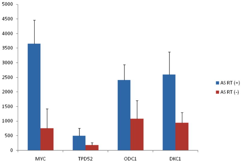
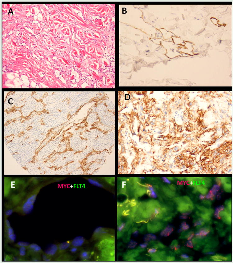
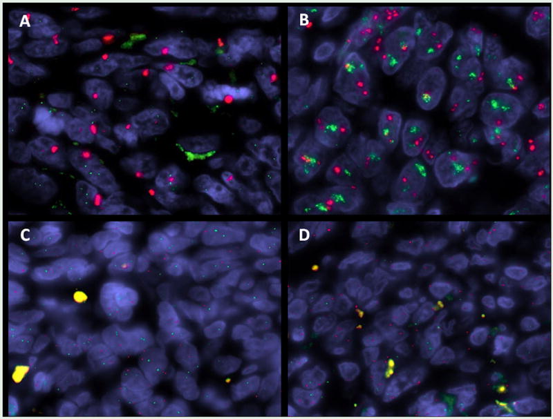
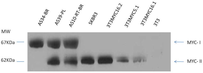
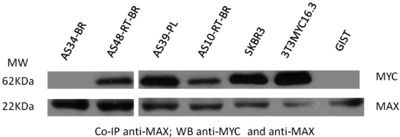
Angiosarcoma (AS) is a distinct group of sarcomas characterized by upregulation of vascular-specific receptor tyrosine kinases, including TIE1, KDR, TEK, and FLT1. In keeping with the clinical heterogeneity, gene-expression profiling distinguishes two AS genomic clusters, which correlate with anatomical location and prior exposure to radiation. Furthermore, a high percentage of secondary AS, but not primary AS, shows distinct 8q24 chromosomal gains, due to MYC amplification. In this study, we mined the transcriptional output of 10 secondary and 11 primary AS to better define the dichotomy in the pathogenesis of these two clinical subsets. The oncogenic role of MYC was investigated further in secondary AS as well as in radiation-induced atypical vascular lesions (AVL) and other radiation-associated sarcomas. High-level MYC amplification was found in 100% of secondary AS, but in none of the AVL or other radiation-associated sarcomas. Coamplification of FLT4 (encoding VEGFR3) was identified in 25% of secondary AS, but not in other types. Our findings reinforce the distinct pathogenesis of AS subtypes, with MYC amplification being an early, but necessary event in secondary AS. Secondary genetic hits, such as FLT4 gene coamplification or KDR mutations, may play a role in tumor progression as well as potential therapeutic targeting.
© 2010 Wiley-Liss, Inc.
Figures
References
-
- Antonescu CR, Besmer P, Guo T, Arkun K, Hom G, Koryotowski B, Leversha MA, Jeffrey PD, Desantis D, Singer S, Brennan MF, Maki RG, DeMatteo RP. Acquired resistance to imatinib in gastrointestinal stromal tumor occurs through secondary gene mutation. Clin Cancer Res. 2005;11:4182–4190. - PubMed
-
- Blackwood MJ. Utilization management and data acquisition: a case study. Benefits Q. 1994;10:38–42. - PubMed
-
- Choschzick M, Lassen P, Lebeau A, Marx AH, Terracciano L, Heilenkotter U, Jaenicke F, Bokemeyer C, Izbicki J, Sauter G, Simon R. Amplification of 8q21 in breast cancer is independent of MYC and associated with poor patient outcome. Mod Pathol. 2010;23:603–610. - PubMed
Publication types
MeSH terms
Substances
Grants and funding
LinkOut - more resources
Full Text Sources
Other Literature Sources
Medical
Research Materials
Miscellaneous
