. 2011 Jan;32(1):33-43.
doi: 10.1002/humu.21377. Epub 2010 Dec 9.

Germline KRAS mutations cause aberrant biochemical and physical properties leading to developmental disorders

Affiliations

Germline KRAS mutations cause aberrant biochemical and physical properties leading to developmental disorders

Lothar Gremer et al. Hum Mutat. 2011 Jan.

Abstract

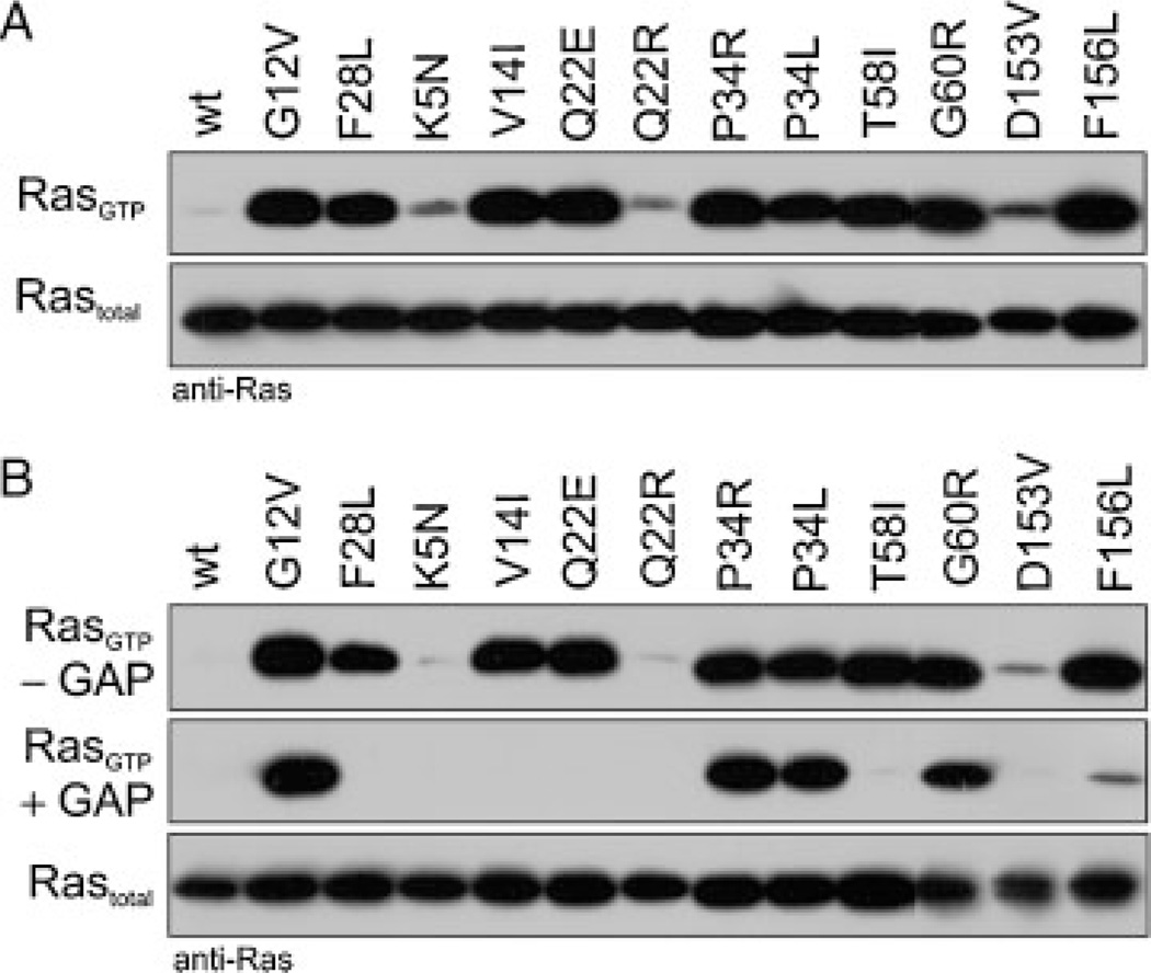
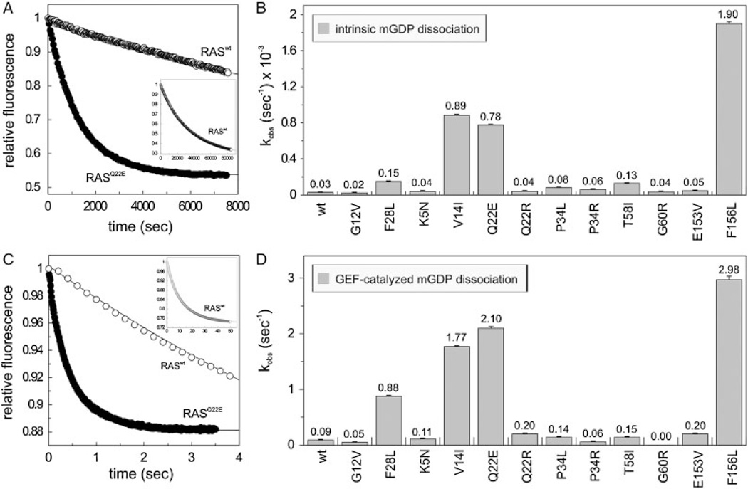
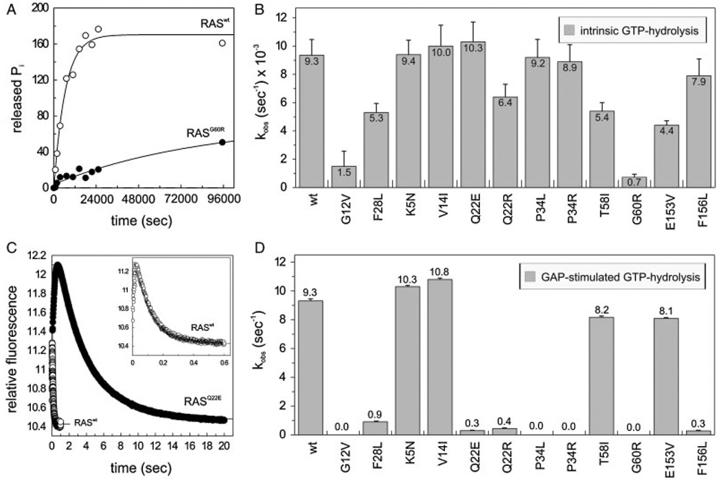
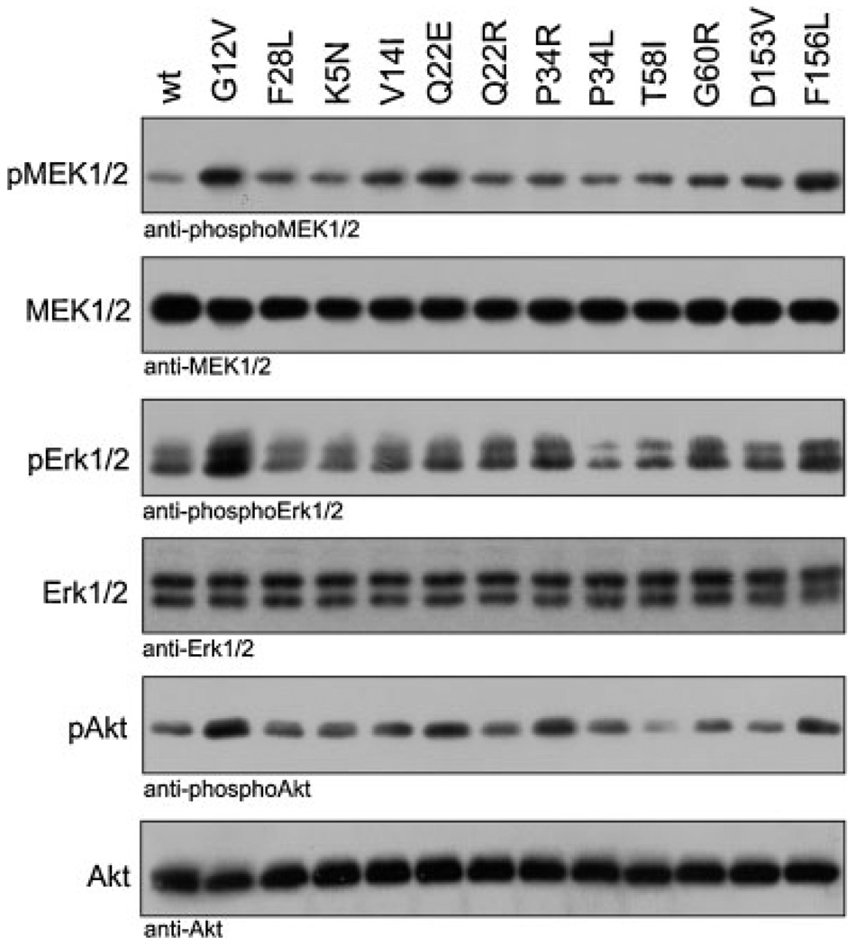
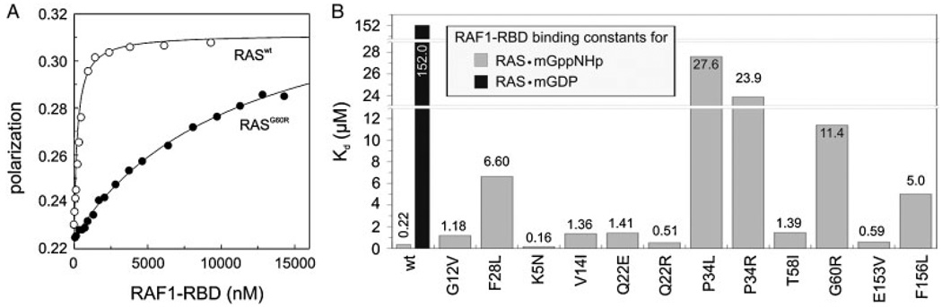
The KRAS gene is the most common locus for somatic gain-of-function mutations in human cancer. Germline KRAS mutations were shown recently to be associated with developmental disorders, including Noonan syndrome (NS), cardio-facio-cutaneous syndrome (CFCS), and Costello syndrome (CS). The molecular basis of this broad phenotypic variability has in part remained elusive so far. Here, we comprehensively analyzed the biochemical and structural features of ten germline KRAS mutations using physical and cellular biochemistry. According to their distinct biochemical and structural alterations, the mutants can be grouped into five distinct classes, four of which markedly differ from RAS oncoproteins. Investigated functional alterations comprise the enhancement of intrinsic and guanine nucleotide exchange factor (GEF) catalyzed nucleotide exchange, which is alternatively accompanied by an impaired GTPase-activating protein (GAP) stimulated GTP hydrolysis, an overall loss of functional properties, and a deficiency in effector interaction. In conclusion, our data underscore the important role of RAS in the pathogenesis of the group of related disorders including NS, CFCS, and CS, and provide clues to the high phenotypic variability of patients with germline KRAS mutations.


Figures

Figure 1
Figure 1
Relative positions of amino acids in KRAS altered in patients with NS, CFCS, and CS. A: Secondary structure elements and conserved motifs of RAS. The α-helices and β-strands are illustrated as cylinders and arrows, respectively. The G-domain of RAS also consists of five conserved motifs (G1–G5; gray boxes) that are responsible for specific and tight nucleotide binding and hydrolysis. Bold lines indicate the position of specific RAS signatures including the hypervariable region (HVR), which is polybasic in KRAS 4B. Amino acids investigated in this study are indicated by arrows. The isoprenylation site of the protein is at the cysteine of the C-terminal CaaX motif. B, C: Solvent accessible surfaces of HRAS molecules are shown in the inactive GDP-bound state (B) and the active GTP-bound state (C). For clarity, structures are illustrated in three different views. Therefore, central panels are rotated 90° around the vertical axes to the right (left panel) and to the left (right panel). Amino acids altered in patients with NS, CFCS, or CS are color coded. Dashed arrows depict critical residues buried within the hydrophobic core of the protein.
Figure 2
Figure 2
Cellular levels of the active, GTP-bound forms of germline KRAS mutants. Pull-down experiments of GTP-bound KRAS proteins (RASGTP) were performed in COS-7 cells transiently expressing either KRASwt or germline KRAS mutants in the presence (A) and in the absence of serum (B). Irrespective of culture conditions almost all KRAS mutants showed an increased GTP-bound level. Purified RAS-GAP, which was added to the cleared cell lysates proved the GAP sensitivity of the mutants (B, middle panel). GAP resistant mutants, RASP34L, RASP34R, and RASG60R, resided in the active state comparable to oncogenic RASG12V. Total amounts of recombinant RAS are shown for equal expression and loading. Anti-RAS antibodies used in these experiments were anti-RAS (RAS10 clone, Upstate-Millipore™, mutants p.G60R, p.D153V, p.F156L) and anti-RAS (BD Transduction Laboratories™, wt and all other mutants), because some mutations modified the RAS epitope recognized by the respective antibodies. Additional information is given in Supp. Fig. S2.
Figure 3
Figure 3
Modified nucleotide exchange properties of the RAS mutants. Intrinsic (A, B) and GEF-catalyzed (C, D) mantGDP dissociation from the RAS proteins (0.2 µM) in the presence of 40 µM GDP (A) or of 40 µM GDP and 2 µM CDC25 (C). On the panels A and C the respective time-dependent reactions of RASwt and a representative RAS mutant (p.Q22E) are shown. On the panels B and D the observed rate constant (kobs) of all RAS proteins are illustrated. RASwt, RASG12V, and RASF28L were included as controls. The insets (in A and C) show the complete time course of the mantGDP dissociation from RASwt. Standard errors of five to seven independent measurements are shown.
Figure 4
Figure 4
GAP insensitivity of the RAS mutants. (A, B) Intrinsic γ32P-GTP hydrolysis reaction rates were measured for individual RAS proteins (1 µM). (C, D) GAP-stimulated GTPase reaction of the RAS proteins (0.2 µM) was measured in the presence of 2 µM NF1–333. On panels A and C, the respective time-dependent reactions of RASwt and a representative RAS mutant (p.G60R in A, p.Q22E in C) are shown. RASwt, RASG12V, and RASF28L were included as controls. The inset in panel C shows the complete and more detailed time course of GAP activity on RASwt. Standard errors of five to seven independent measurements are shown.
Figure 5
Figure 5
Increased downstream signaling activity of the germline RAS mutants under serum-free conditions. KRAS mutants, transiently transfected in COS-7 cells were analyzed for the phosphorylation level of MEK1/2 (pMEK1/2), ERK1/2 (pERK1/2), and AKT (pAKT) under serum-free culture conditions. The amounts of total RAS, MEK1/2, ERK1/2, and AKT in the cleared cell lysates as well as RASwt, RASG12V, and RASF28L were included as controls. Results of experiments in the presence of serum are shown in Supp. Fig. S4.
Figure 6
Figure 6
Significant loss of RAF1 binding affinity for RAS mutants. Binding of RAF1-RBD (increasing concentrations as indicated) to mantGppNHp-bound RAS (0.2 µM) was measured using fluorescence polarization. On the panel A, the respective concentration-dependent measurements of RASwt and a representative RAS mutant (p.G60R) are shown. On panel B, the dissociation constants (Kd) of all RAS proteins are illustrated. RASwt, RASG12V, and RASF28L were included as controls. Data obtained with another RAS effector, RALGDS are shown in Supp. Fig. S5.

References

    1. Abankwa D, Hanzal-Bayer M, Ariotti N, Plowman SJ, Gorfe AA, Parton RG, McCammon JA, Hancock JF. A novel switch region regulates H-ras membrane orientation and signal output. EMBO J. 2008;27:727–735. - PMC - PubMed
    1. Ahmadian MR. Prospects for anti-ras drugs. Br J Haematol. 2002;116:511–518. - PubMed
    1. Ahmadian MR, Hoffmann U, Goody RS, Wittinghofer A. Individual rate constants for the interaction of Ras proteins with GTPase-activating proteins determined by fluorescence spectroscopy. Biochemistry. 1997;36:4535–4541. - PubMed
    1. Ahmadian MR, Wittinghofer A, Herrmann C. Fluorescence methods in the study of small GTP-binding proteins. Methods Mol Biol. 2002;189:45–63. - PubMed
    1. Ahmadian MR, Zor T, Vogt D, Kabsch W, Selinger Z, Wittinghofer A, Scheffzek K. Guanosine triphosphatase stimulation of oncogenic Ras mutants. Proc Natl Acad Sci USA. 1999;96:7065–7070. - PMC - PubMed

Publication types

MeSH terms

Supplementary concepts