Comparative Study
. 2010 Nov 23;49(46):9948-56.
doi: 10.1021/bi101165p. Epub 2010 Nov 1.

Characterization and comparison of two binding sites on obscurin for small ankyrin 1

Affiliations
Comparative Study

Characterization and comparison of two binding sites on obscurin for small ankyrin 1

Ben Busby et al. Biochemistry. .

Abstract

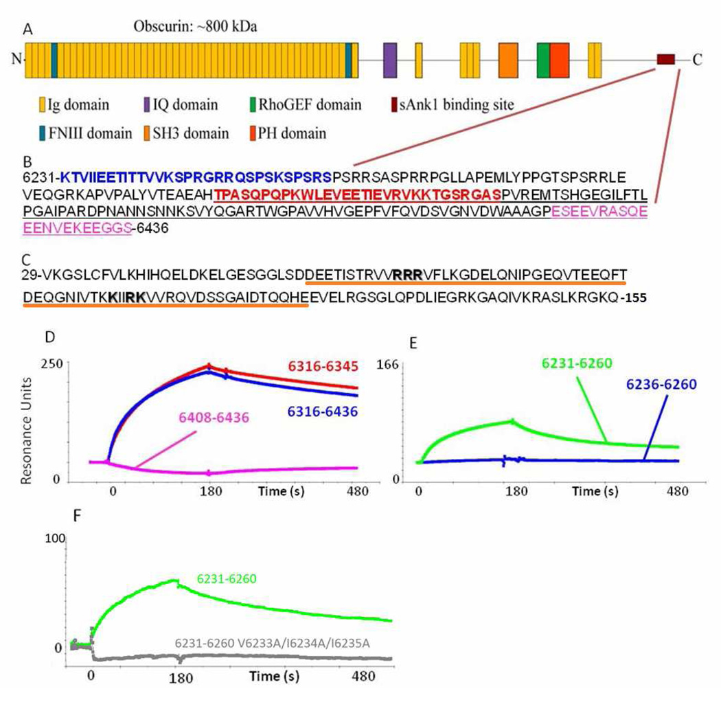
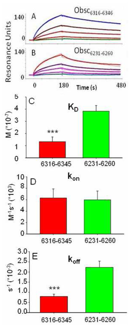
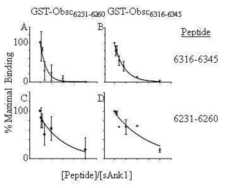
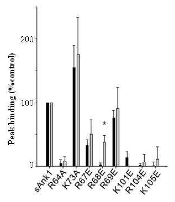
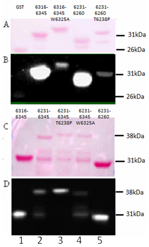
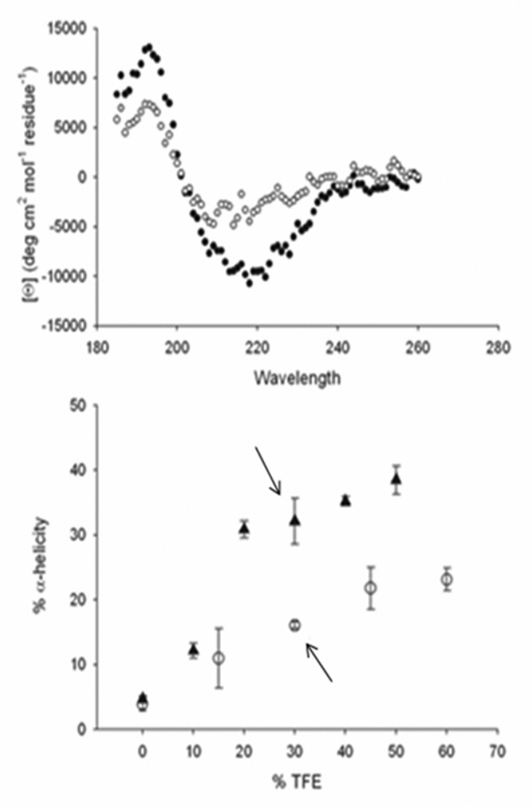
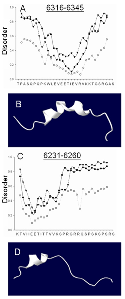
Obscurin A, an ∼720 kDa modular protein of striated muscles, binds to small ankyrin 1 (sAnk1, Ank 1.5), an integral protein of the sarcoplasmic reticulum, through two distinct carboxy-terminal sequences, Obsc(6316-6436) and Obsc(6236-6260). We hypothesized that these sequences differ in affinity but that they compete for the same binding site on sAnk1. We show that the sequence within Obsc(6316-6436) that binds to sAnk1 is limited to residues 6316-6345. Comparison of Obsc(6231-6260) to Obsc(6316-6345) reveals that Obsc(6316-6345) binds sAnk1 with an affinity (133 ± 43 nM) comparable to that of the Obsc(6316-6436) fusion protein, whereas Obsc(6231-6260) binds with lower affinity (384 ± 53 nM). Oligopeptides of each sequence compete for binding with both sites at half-maximal inhibitory concentrations consistent with the affinities measured directly. Five of six site-directed mutants of sAnk1 showed similar reductions in binding to each binding site on obscurin, suggesting that they dock to many of the same residues of sAnk1. Circular dichroism (CD) analysis of the synthetic oligopeptides revealed a 2-fold greater α-helical content in Obsc(6316-6346), ∼35%, than Obsc(6231-6260,) ∼17%. Using these data, structural prediction algorithms, and homology modeling, we predict that Obsc(6316-6345) contains a bent α-helix of 12 amino acids, flanked by short disordered regions, and that Obsc(6231-6260) has a short, N-terminal α-helix of 4-5 residues followed by a long disordered region. Our results are consistent with a model in which both sequences of obscurin differ significantly in structure but bind to the ankyrin-like repeat motifs of sAnk1 in a similar though not identical manner.


Figures

Fig. 1
Fig. 1. The sequences of obscurin and sAnk1 that mediate binding
(A) Cartoon of the domain organization of obscurin A. The burgundy lines indicate the region of the molecule shown in B. (B) The sequence of residues corresponding to amino acids 6231–6436 of obscurin A (Rattus norveigicus). Blue text denotes the sequence identified by Bagnato et al.. The red sequence denotes the residues identified as the minimal binding domain and used for experiments in this manuscript (see D and E). The underlined sequence is that identified by Kontrogianni-Konstantopoulos et al.. The sequence in pink shows a sequence rich in electronegative amino acids that is neither necessary nor sufficient to bind sAnk1 (see D, pink). (C) Sequence of residues 29–155 of sAnk1 of the rat. Residues shown by Borzok et al. to mediate binding to obscurin are shown in bold; residues 57–122, underlined in orange have previously been modeled as ankyrin-like repeats. (D, E) Surface plasmon resonance assays of binding of fusion constructs of sAnk129–155 to different sequences of obscurin. (D) Obsc6316–6345 (red), Obsc6316–6436 (blue), Obsc6408–6436 (pink). (E) Obsc6231–6260 (green), Obsc6236–6260 (blue). (F) Obsc6231–6260 (green), Obsc6231–6260 V6233A/I6234A/I6235A (grey).
Fig. 2
Fig. 2. Kinetics of binding of GST-Obsc6231–6260 and GST-Obsc6316–6345 to MBP-sAnk129–155
(A, B) Colored curves show binding of serial diluted concentrations of MBP-sAnk1 starting at 3µM to GST-Obsc6316–6345 (A) and GST-Obsc6231–6260 (B). Black curves are fits based on a 1:1 stoichiometry of binding. (C–E) Bar graphs of values of KD, kon, and koff for GST-Obsc6316–6345 A (red) and GST-Obsc6231–6260 (green). *** denotes p < .05. n = 5 for all values.
Fig. 3
Fig. 3. Inhibition of binding of sAnk1 to GST-Obsc6316–6345 and GST-Obsc6231–6260 by synthetic oligopeptides
Solutions containing 1µM MBP-sAnk1 were pre-mixed with fusion proteins containing each of the binding sites of obscurin with different relative molar amounts (shown on ordinate axis) of synthetic oligopeptide of either Obsc6316–6345 or Obsc6231–6260. (A, B) Synthetic oligopeptide 6316–6345 inhibits binding of MBP-sAnk129–155 to GST-Obsc6231–6260 (A) and GST-Obsc6316–6345 (B). (C, D) Synthetic oligopeptide 6231–6260 inhibits binding of MBP-sAnk129–155 to both GST-Obsc6231–6260 (C) and GST-Obsc6316–6345 (D) but at ratios significantly higher than oligopeptide 6316–6345. For all experiments with oligopeptide 6231–6260 experiments, n=5; for experiments with oligopeptide 6316–6345, n=6, except for those at 10:1 and 20:1 ratios, for which n=4.
Fig. 4
Fig. 4. Site-directed mutants of sAnk1 reduce binding of Obsc6231–6260 and Obsc6316–6345 to a similar extent
We assayed the effects on binding of mutating the lysine or arginine residues of sAnk1 involved in binding obscurin (see Fig. 1) to glutamates or alanines. Grey bars: Binding to Obsc6231–6260 (normalized to maximal binding, measured with WT sAnk1); black bars: binding to Obsc6316–6345 (normalized similarly). With the exception of sAnk1 R68E, mutations in sAnk1 have similar effects on binding to each of the binding sites on obscurin. n=5 for all experiments. Error bars, S.D.; * indicates a significant difference (p < .05).
Fig. 5
Fig. 5. Residues 6316–6345 of obscurin dominate binding to sAnk1 when both binding sites are present
All GST constructs following transfer to nitrocellulose are shown in panel A and C with a Ponceau Red stain, which show equal loading. Panels B and D show the results of overlay assays of these nitrocellulose blots. Mutations of W6325 of Obsc6316–6345 to alanine (A,B; Lane 3, GST-Obsc6316–6345 W6325A) and T6328 of Obsc6231–6260 to proline (A,B; Lane 5, GST-Obsc6231–6260T6328P) completely ablate binding of the individual sites to MBP-sAnk129–155 in blot overlays and are barely greater than GST vector alone (A,B; Lane 1). Binding of WT constructs are shown in the adjacent lanes: Lane 2 (GST-Obsc6316–6345), Lane 4 (GST-Obsc6231–6260). These bands run at 31–32 kDa, consistent with the size of the GST fusion constructs. This developed blot in panel B was intentionally overexposed to show the dramatic nature of the inhibition by the mutants which is completely undetectable at low exposures. The GST-Obsc6231–6345 construct, which contains both binding sequences, is difficult to purify and is unstable, so only a small amount runs at the appropriate molecular mass of 38 kDa (C,D; Lane 2); a breakdown product, which retains the binding site within residues 6231–6260, is present at 31 kDa and also binds MBP-sAnk129–155. Mutation of W6325 of GST-Obsc6231–6345 to A (Lane 4: GST-Obsc6231–6345W6325A) reduces binding of the 38kDa band to MBP-sAnk1 almost completely (compare to Lane 2). Mutation of T6328 of GST-Obsc6231–6345 to P (lane 3, GST-Obsc6231–6345T6328P) does not inhibit binding of the 38kDa band to MBP-sAnk129–155 and may in fact enhance it (compared to Lanes 2 and 4). The 31 kDa breakdown product of GST-Obsc6231–6345T6328P does not bind to MBP-sAnk129–155, however, consistent with the effects of this mutation on GST-Obsc6231–6260. Both individual sites, GST-Obsc6316–6345 (Lane 1) and GST-Obsc6231–6260 (Lane 5), like panels A and B bind MBP-sAnk1. This experiment was repeated 5 times, with the same results.
Fig. 6
Fig. 6. CD spectra of synthetic oligopeptides of Obsc6231–6260 and Obsc6316–6345
(A) The CD spectra of both peptides in the presence of 30% TFE are represented by triangles (6316–6345) and open circles (6231–6260) Obsc6316–6345 shows a greater degree of α-helicity, with deeper minima at ~220nm and ~208nm. (B) α-Helicity of the oligopeptides as a function of TFE concentration. Arrows indicate α-helicity at 30% TFE. n = 4.
Fig. 7
Fig. 7. Disorder plots and structural models of Obsc6231–6260 and Obsc6316–6345
(A, C) Disorder plots: circles represent the predicted mobility of α-carbons in the oligopeptide sequences of obscurin at 1000 K; triangles represent regions for which similar short oligopeptide sequences have never been experimentally solved; closed circles represent predicted random coiled domains, a necessary but not sufficient condition for disorder. (B, D) Homology model for Obsc6316–6345 (based on RelA) and a representation of Obsc6231–6260, based on relative alpha helicity and overall similarity to known ankyrin binding motifs such as CDK4, both in good agreement with CD data (Fig. 6) disorder plots (this figure) and secondary structure prediction algorithms (not shown).

Similar articles

Cited by

References

    1. Zhou D, Birkenmeier CS, Williams MW, Sharp JJ, Barker JE, Bloch RJ. Small, membrane-bound, alternatively spliced forms of ankyrin 1 associated with the sarcoplasmic reticulum of mammalian skeletal muscle. J. Cell Biol. 1997;136:621–631. - PMC - PubMed
    1. Birkenmeier CS, Sharp JJ, Gifford EJ, Deveau SA, Barker JE. An alternative first exon in the distal end of the erythroid ankyrin gene leads to production of a small isoform containing an NH2-terminal membrane anchor. Genomics. 1998;50:79–88. - PubMed
    1. Bennett V, Baines AJ. Spectrin and ankyrin-based pathways: metazoan inventions for integrating cells into tissues. Physiol Rev. 2001;81:1353–1392. - PubMed
    1. Bowman AL, Kontrogianni-Konstantopoulos A, Hirsch SS, Geisler SB, Gonzalez-Serratos H, Russell MW, Bloch RJ. Different obscurin isoforms localize to distinct sites at sarcomeres. FEBS Lett. 2007;581:1549–1554. - PMC - PubMed
    1. Carlsson L, Yu JG, Thornell LE. New aspects of obscurin in human striated muscles. Histochem. Cell Biol. 2008;130:91–103. - PubMed

Publication types