. 2010 Oct 15:10:261.
doi: 10.1186/1471-2180-10-261.

Planctomycetes dominate biofilms on surfaces of the kelp Laminaria hyperborea

Affiliations

Planctomycetes dominate biofilms on surfaces of the kelp Laminaria hyperborea

Mia M Bengtsson et al. BMC Microbiol. .

Abstract

Background: Bacteria belonging to Planctomycetes display several unique morphological and genetic features and are found in a wide variety of habitats on earth. Their ecological roles in these habitats are still poorly understood. Planctomycetes have previously been detected throughout the year on surfaces of the kelp Laminaria hyperborea from southwestern Norway. We aimed to make a detailed investigation of the abundance and phylogenetic diversity of planctomycetes inhabiting these kelp surfaces.

Results: Planctomycetes accounted for 51-53% of the bacterial biofilm cells in July and September and 24% in February according to fluorescence in situ hybridization (FISH) results. Several separate planctomycetes lineages within Pirellulae, Planctomyces and OM190 were represented in 16S rRNA gene clone libraries and the most abundant clones belonged to yet uncultured lineages. In contrast to the abundance, the diversity of the planctomycete populations increased from July to February and was probably influenced by the aging of the kelp tissue. One planctomycete strain that was closely related to Rhodopirellula baltica was isolated using selective cultivation techniques.

Conclusions: Biofilms on surfaces of L. hyperborea display an even higher proportion of planctomycetes compared to other investigated planctomycete-rich habitats such as open water, sandy sediments and peat bogs. The findings agree well with the hypothesis of the role of planctomycetes as degraders of sulfated polymeric carbon in the marine environment as kelps produce such substances. In addition, the abundant planctomycete populations on kelp surfaces and in association with other eukaryotes suggest that coexistence with eukaryotes may be a key feature of many planctomycete lifestyles.


Figures
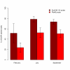
Figure 1
Figure 1
Abundance of planctomycetes in kelp surface biofilms. The abundance of cells stained by the Planctomycetes specific probe Pla46 and the general bacterial probe Eub338 I-III at three different sampling times as a percentage of total cells (DAPI stained). The height of the bars represents the average percentage values of six individual kelp plants sampled at each sampling occasion. Error bars indicate one standard deviation (± 1SD).
Figure 2
Figure 2
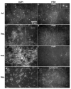
Distribution of planctomycete cells in the biofilm. Fluorescence microscopy images of Laminaria hyperborea surface biofilm. Images a, c, e and g show DAPI stained biofilm while b, d, f and h show FISH signals in the same microscope fields from hybridizations with either the Pla46 probe (b, d and f) or the Eub 338 I-III probe mix (h). Images show representative microscope fields of samples from July 2007 (a-b), September 2008 (c-d, g-h) and February 2007 (e-f). The enlarged inset image in b shows the typical ring shaped FISH signals of planctomycetes.
Figure 3
Figure 3
The P1 strain. A phase contrast photomicrograph showing the Rhodopirellula sp. strain P1 isolated from kelp surface biofilm, displaying ovoid cells, budding and rosette formation.
Figure 4
Figure 4
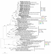
Phylogenetic relationships of planctomycetes. A maximum likelihood (PhyML) tree based on 16S sequences of Planctomycetes. An outgroup consisting of reference sequences from the Verrucomicrobia were used for tree calculation, but is not displayed in the tree. Bold letters designate sequences derived from the present study, which include one representative of each OTU and the P1 isolate. Reference sequences from the SILVA database are described by their GenBank accession numbers, origin of the sequence (environmental or cultured strain) and the habitat they were obtained from. The vertical lines mark phylogenetic lineages of interest. The percentage of each clone library that was made up of sequences from each phylogenetic lineage is indicated. Bootstrap values >60 (based on 1000 bootstraps) are displayed. The scale bar indicates 0.10 (10%) sequence divergence.
Figure 5
Figure 5
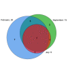
Overlap of planctomycete OTUs between sampling times. A Venn diagram describing the degree of OTU overlap between the different clone libraries. The total number of OTUs in each library is displayed outside the circles and the number of overlapping OTUs is given inside the areas of the circles. The area-proportional Venn diagram was generated at http://www.venndiagram.tk.
Figure 6
Figure 6
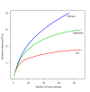
OTU diversity of planctomycetes. Rarefaction curves indicating the expected OTU richness of the clone libraries with different sampling efforts.

References

    1. Lindsay MR, Webb RI, Strous M, Jetten MS, Butler MK, Forde RJ, Fuerst JA. Cell compartmentalisation in planctomycetes: novel types of structural organisation for the bacterial cell. Arch Microbiol. 2001;175(6):413–429. doi: 10.1007/s002030100280. - DOI - PubMed
    1. Fuerst JA. Intracellular compartmentation in planctomycetes. Annual review of microbiology. 2005;59:299–328. doi: 10.1146/annurev.micro.59.030804.121258. - DOI - PubMed
    1. Strous M, Fuerst JA, Kramer EHM, Logemann S, Muyzer G, van de Pas-Schoonen KT, Webb R, Kuenen JG, Jetten MSM. Missing lithotroph identified as new planctomycete. Nature. 1999;400(6743):446–449. doi: 10.1038/22749. - DOI - PubMed
    1. Ward N, Staley JT, Fuerst JA, Giovannoni S, Schlesner H, Stackebrandt E. The Prokaryotes. 3. Vol. 7. New York: Springer; 2006. The Order Planctomycetales, Including the Genera Planctomyces, Pirellula, Gemmata and Isosphaera and the Candidatus Genera Brocadia, Kuenenia and Scalindua.
    1. Delong EF, Franks DG, Alldredge AL. Phylogenetic diversity of aggregate-attached vs. free-living marine bacterial assemblages. Limnol Oceanogr. 1993;38(5):924–934. doi: 10.4319/lo.1993.38.5.0924. - DOI

Publication types

LinkOut - more resources