

Abnormal structure or function of the amygdala is a common component of neurodevelopmental disorders
- PMID: 20950634
- PMCID: PMC3060967
- DOI: 10.1016/j.neuropsychologia.2010.09.028
Abnormal structure or function of the amygdala is a common component of neurodevelopmental disorders
Abstract
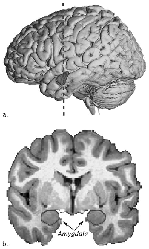
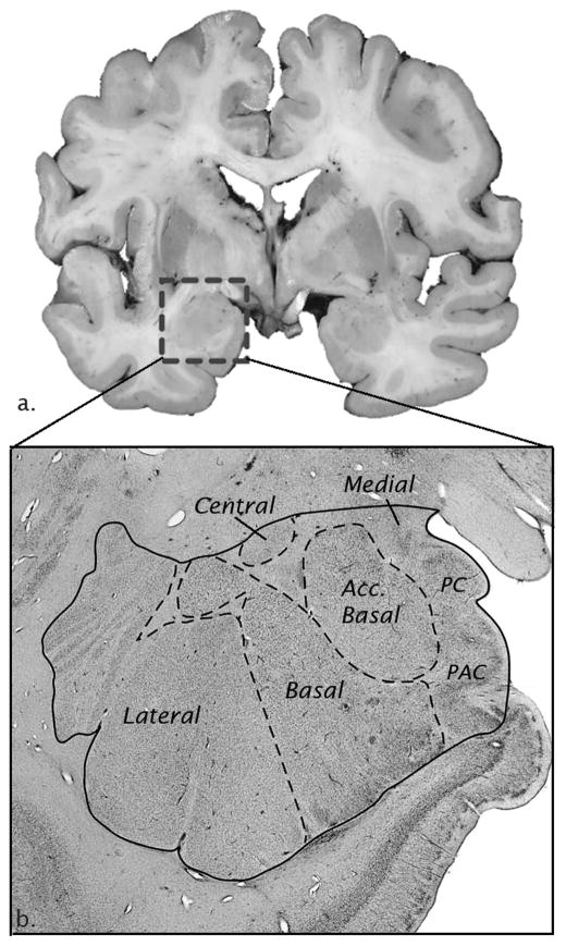
The amygdala, perhaps more than any other brain region, has been implicated in numerous neuropsychiatric and neurodevelopmental disorders. It is part of a system initially evolved to detect dangers in the environment and modulate subsequent responses, which can profoundly influence human behavior. If its threshold is set too low, normally benign aspects of the environment are perceived as dangers, interactions are limited, and anxiety may arise. If set too high, risk taking increases and inappropriate sociality may occur. Given that many neurodevelopmental disorders involve too little or too much anxiety or too little of too much social interaction, it is not surprising that the amygdala has been implicated in many of them. In this chapter, we begin by providing a brief overview of the phylogeny, ontogeny, and function of the amygdala and then appraise data from neurodevelopmental disorders which suggest amygdala dysregulation. We focus on neurodevelopmental disorders where there is evidence of amygdala dysregulation from postmortem studies, structural MRI analyses or functional MRI. However, the results are often disparate and it is not totally clear whether this is due to inherent heterogeneity or differences in methodology. Nonetheless, the amygdala is a common site for neuropathology in neurodevelopmental disorders and is therefore a potential target for therapeutics to alleviate associated symptoms.
Published by Elsevier Ltd.
Figures
References
-
- Adolphs R, Gosselin F, Buchanan TW, Tranel D, Schyns P, Damasio AR. A mechanism for impaired fear recognition after amygdala damage. Nature. 2005;433(7021):68–72. - PubMed
Publication types
MeSH terms
Grants and funding
LinkOut - more resources
Full Text Sources
Medical
