Review
. 2011 Apr;1813(4):597-607.
doi: 10.1016/j.bbamcr.2010.10.008. Epub 2010 Oct 13.

Mitochondrial involvement in cell death of non-mammalian eukaryotes

Affiliations
Review

Mitochondrial involvement in cell death of non-mammalian eukaryotes

Eltyeb Abdelwahid et al. Biochim Biophys Acta. 2011 Apr.

Abstract

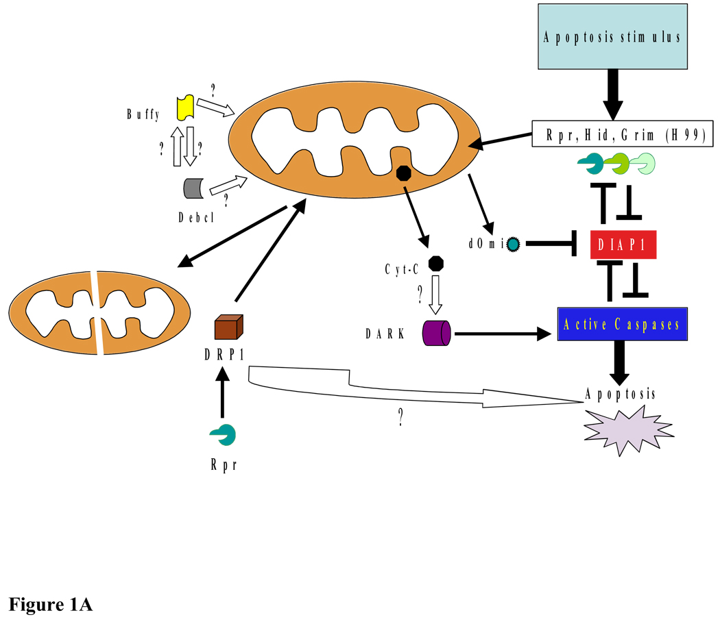
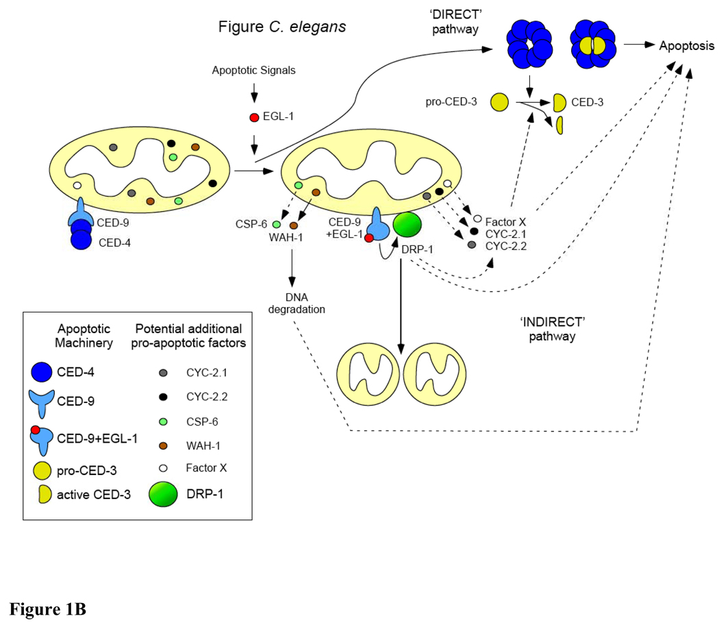
Although mitochondria are essential organelles for long-term survival of eukaryotic cells, recent discoveries in biochemistry and genetics have advanced our understanding of the requirements for mitochondria in cell death. Much of what we understand about cell death is based on the identification of conserved cell death genes in Drosophila melanogaster and Caenorhabditis elegans. However, the role of mitochondria in cell death in these models has been much less clear. Considering the active role that mitochondria play in apoptosis in mammalian cells, the mitochondrial contribution to cell death in non-mammalian systems has been an area of active investigation. In this article, we review the current research on this topic in three non-mammalian models, C. elegans, Drosophila, and Saccharomyces cerevisiae. In addition, we discuss how non-mammalian models have provided important insight into the mechanisms of human disease as they relate to the mitochondrial pathway of cell death. The unique perspective derived from each of these model systems provides a more complete understanding of mitochondria in programmed cell death. This article is part of a Special Issue entitled Mitochondria: the deadly organelle.


Figures

Figure 1
Figure 1
A: Mitochondria and apoptosis in Drosophila. Cell death in Drosophila is activated by Rpr, Hid, Grim, and Skl, which bind to DIAP1 and inhibit DIAP1’s caspase inhibitory function. The role of cytochrome c in Drosophila caspase activation is not well characterized, however, a role for the mitochondria in the apoptotic process is suggested by the several findings, including Rpr, Grim, and Hid localization to the mitochondria. On initiation of apoptosis, Rpr and Hid are transcribed and rapidly localize to mitochondria, resulting in changes in mitochondrial ultrastructure and Cyt-c release. Drp 1-dependent mitochondrial disruption appears to be required for Drosophila apoptosis. Upon apoptosis induction, dOmi is released into the cytosol, where it binds DIAP1 causing caspase activation. The role of Drosophila Bcl-2-like proteins Buffy and Debcl in fly apoptosis remains unclear. Additionally, despite a requirement of Dark for caspase activation, the role of Drosophila apoptosome in caspase activation is not well understood. B: Mitochondria and apoptosis in C. elegans. Two apoptotic pathways are at work in C. elegans. During apoptosis, the pro-apoptotic BH3-only protein EGL-1 binds to the anti-apoptotic Bcl-2-like protein CED-9, thereby releasing the pro-apoptotic Apaf-1 like protein CED-4. Released CED-4 oligomerizes and promotes the processing of pro-caspase CED-3. CED-4 and active CED-3 form a holoenzyme that triggers apoptosis (‘DIRECT’ pathway). In a parallel pathway, when bound to EGL-1, CED-9 adopts a pro-apoptotic conformation and activates the dynamin-related protein DRP1. Active DRP1 promotes mitochondrial fragmentation and apoptosis by enhancing CED-3 processing or by acting in parallel to CED-3. The pro-apoptotic role of DRP1 could involve the release of potential pro-apoptotic factors (such as Cyt-c (CYC-2.1 or CYC-2.2) or an unknown factor X) but seems to be unrelated to its role in mitochondrial fission (‘INDIRECT’ pathway). Later during apoptosis, the endonuclease CSP-6 and the AIF homolog WAH-1 are released from mitochondria to degrade nuclear DNA, thereby participating in the dismantlement of the cell.
Figure 1
Figure 1
A: Mitochondria and apoptosis in Drosophila. Cell death in Drosophila is activated by Rpr, Hid, Grim, and Skl, which bind to DIAP1 and inhibit DIAP1’s caspase inhibitory function. The role of cytochrome c in Drosophila caspase activation is not well characterized, however, a role for the mitochondria in the apoptotic process is suggested by the several findings, including Rpr, Grim, and Hid localization to the mitochondria. On initiation of apoptosis, Rpr and Hid are transcribed and rapidly localize to mitochondria, resulting in changes in mitochondrial ultrastructure and Cyt-c release. Drp 1-dependent mitochondrial disruption appears to be required for Drosophila apoptosis. Upon apoptosis induction, dOmi is released into the cytosol, where it binds DIAP1 causing caspase activation. The role of Drosophila Bcl-2-like proteins Buffy and Debcl in fly apoptosis remains unclear. Additionally, despite a requirement of Dark for caspase activation, the role of Drosophila apoptosome in caspase activation is not well understood. B: Mitochondria and apoptosis in C. elegans. Two apoptotic pathways are at work in C. elegans. During apoptosis, the pro-apoptotic BH3-only protein EGL-1 binds to the anti-apoptotic Bcl-2-like protein CED-9, thereby releasing the pro-apoptotic Apaf-1 like protein CED-4. Released CED-4 oligomerizes and promotes the processing of pro-caspase CED-3. CED-4 and active CED-3 form a holoenzyme that triggers apoptosis (‘DIRECT’ pathway). In a parallel pathway, when bound to EGL-1, CED-9 adopts a pro-apoptotic conformation and activates the dynamin-related protein DRP1. Active DRP1 promotes mitochondrial fragmentation and apoptosis by enhancing CED-3 processing or by acting in parallel to CED-3. The pro-apoptotic role of DRP1 could involve the release of potential pro-apoptotic factors (such as Cyt-c (CYC-2.1 or CYC-2.2) or an unknown factor X) but seems to be unrelated to its role in mitochondrial fission (‘INDIRECT’ pathway). Later during apoptosis, the endonuclease CSP-6 and the AIF homolog WAH-1 are released from mitochondria to degrade nuclear DNA, thereby participating in the dismantlement of the cell.

References

    1. Yuan J, Shaham S, Ledoux S, Ellis HM, Horvitz HR. The C. elegans cell death gene ced-3 encodes a protein similar to mammalian interleukin-1 beta-converting enzyme. Cell. 1993;75:641–652. - PubMed
    1. Miura M, Zhu H, Rotello R, Hartwieg EA, Yuan J. Induction of apoptosis in fibroblasts by IL-1 beta-converting enzyme, a mammalian homolog of the C. elegans cell death gene ced-3. Cell. 1993;75:653–660. - PubMed
    1. Hengartner MO, Horvitz HR. C. elegans cell survival gene ced-9 encodes a functional homolog of the mammalian proto-oncogene bcl-2. 1994;76:665–676. - PubMed
    1. Crook NE, Clem RJ, Miller LK. An apoptosis-inhibiting baculovirus gene with a zinc finger-like motif. J. Virol. 1993;67:2168–2174. - PMC - PubMed
    1. Hay BA, Wassarman DA, Rubin GM. Drosophila homologs of baculovirus inhibitor of apoptosis proteins function to block cell death. Cell. 1995;83:1253–1262. - PubMed

Publication types

LinkOut - more resources