. 2011 Feb;32(4):1204-17.
doi: 10.1016/j.biomaterials.2010.08.117. Epub 2010 Oct 14.

Doxycycline hydrogels with reversible disulfide crosslinks for dermal wound healing of mustard injuries

Affiliations

Doxycycline hydrogels with reversible disulfide crosslinks for dermal wound healing of mustard injuries

SivaNaga S Anumolu et al. Biomaterials. 2011 Feb.

Abstract

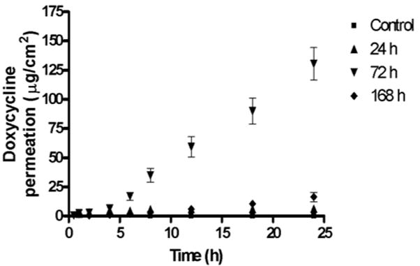
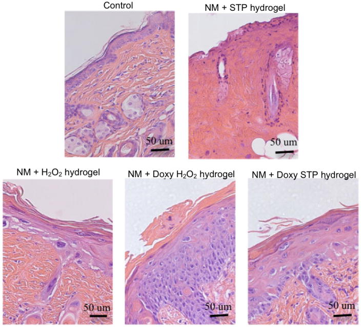
Doxycycline hydrogels containing reversible disulfide crosslinks were investigated for a dermal wound healing application. Nitrogen mustard (NM) was used as a surrogate to mimic the vesicant effects of the chemical warfare agent sulfur mustard. An 8-arm-poly(ethylene glycol) (PEG) polymer containing multiple thiol (-SH) groups was crosslinked using hydrogen peroxide (H(2)O(2) hydrogel) or 8-arm-S-thiopyridyl (S-TP hydrogel) to form a hydrogel in situ. Formulation additives (glycerin, PVP and PEG 600) were found to promote dermal hydrogel retention for up to 24 h. Hydrogels demonstrated high mechanical strength and a low degree of swelling (< 1.5%). Doxycycline release from the hydrogels was biphasic and sustained for up to 10-days in vitro. Doxycycline (8.5 mg/cm(3)) permeability through NM-exposed skin was elevated as compared to non vesicant-treated controls at 24, 72 and 168 h post-exposure with peak permeability at 72 h. The decrease in doxycycline permeability at 168 h correlates to epidermal re-epithelialization and wound healing. Histology studies of skin showed that doxycycline loaded (0.25% w/v) hydrogels provided improved wound healing response on NM-exposed skin as compared to untreated skin and skin treated with placebo hydrogels in an SKH-1 mouse model. In conclusion, PEG-based doxycycline hydrogels are promising for dermal wound healing application of mustard injuries.


Figures

Figure 1
Figure 1
The DSC thermograms show the melting of 8-arm-PEG-SH and 8-arm-PEG-S-TP at 53.37°C and 45.81°C respectively, indicative of conversion of thiols to thiopyridine.
Figure 2
Figure 2
The XPS spectrum for the 8-arm-PEG-S-TP. A) The peak centered at 399eV corresponds to N1s confirming the incorporation of nitrogen in the 8-arm-PEG-S-TP polymer; B) The peak centered at 164eV represents S 2p of sulfur corresponding to the dithiopyridine groups. The XPS analysis of 8-arm-PEG-TP, shows the diffractograms for nitrogen and sulfur atoms. The ratio between sulfur and nitrogen was found to be 2:1.
Figure 3
Figure 3
Effect of the concentration of polymers on swelling kinetics of 4, 8 % H2O2 hydrogels and 5, 8 % S-TP hydrogels. As the concentration of polymers is increased, the degree of swelling is lowered.
Figure 4
Figure 4
Influence of strain (A) and frequency (B) on G′ and G″ of 4 and 8 % H2O2 hydrogels with and without formulation additives. The strain sweep test establishes the regime of linear viscoelasticity (LVE). The frequency sweep test shows that the hydrogels are elastic than viscous and that they have the ability to resist structural changes under strain.
Figure 5
Figure 5
Influence of strain (A) and frequency (B) on G′ and G″ of 5 and 8 % S-TP hydrogels with and without formulation additives. The strain sweep test establishes the regime of linear viscoelasticity (LVE). The frequency sweep test shows that the hydrogels are elastic than viscous and that they have the ability to resist structural changes under strain.
Figure 6
Figure 6
The DSC thermograms for the 4 and 8 % H2O2 hydrogels (A) and 5 and 8 % S-TP hydrogels (B) with and without formulation additives. With an increase in polymer concentration of the hydrogels, the melting of the PEG shifts to lower temperatures. The presence of formulation additives further restricts the motion of PEG chains lowering their melting temperature.
Figure 7
Figure 7
Cumulative amount of doxycycline released as a function of time for 4 and 8 % H2O2 hydrogels and 5 and 8 % S-TP hydrogels. The release data were fitted using a two-phase exponential association equation using GraphPad Prism 4 software. The goodness of fit for the different hydrogels varied from 0.83 to 0.96. The release mechanism for the H2O2 hydrogels is non-fickian or anomalous involving both diffusion and polymer relaxation (0.5<n<1). The release mechanism for the S-TP hydrogels is super case II transport involving relaxation of the polymer as the hydrogel swells (n>1).
Figure 8
Figure 8
Cumulative amount of doxycycline permeated as a function of time through NM-exposed skin. The permeability of doxycycline through NM-exposed skin for variable time periods was found to increase significantly (p<0.01) compared to the control. The order of permeation of doxycycline is 72 h> 168 h> 24 h>control.
Figure 9
Figure 9
Histology of mouse skin 24 h post exposure to NM and hydrogels (Magnification 40x). Two h after topical application of NM or control, either placebo- or doxycycline-containing H2O2 or S-TP hydrogels were applied to the skin. After 24 h, NM was found to cause a marked inflammatory cell infiltration and edema; areas of epidermal/dermal separation were also evident. Epidermal separation was not evident in skin treated with either placebo or doxycycline hydrogels indicating that the hydrogels were acting as an occlusive bandage.
Figure 10
Figure 10
Histology of mouse skin 72 h post exposure to NM and hydrogels (Magnification 40x). NM caused degradation of the dermis, epidermal necrosis and extensive epidermal/dermal separation. The placebo and doxycycline containing H2O2- or S-TP hydrogels suppressed dermal degradation as well as necrosis and blistering. There was marked improvement in the epidermis in doxycycline containing hydrogels. Significant acanthosis, a marker of wound repair, was noted in the skin treated with both doxycycline-containing hydrogels.
Figure 11
Figure 11
Histology of mouse skin 168 h post exposure to NM and hydrogels (Magnification 40x). NM caused extensive necrosis of the skin. Both placebo H2O2- or S-TP hydrogels provided some protection; skin treated with the H2O2-hydrogel retained epidermis, although nuclear degradation was evident. A significant improvement in the skin was found post-treatment with both doxycycline containing hydrogels. This included marked epidermal hyperplasia and hyperkeratosis, which extended into the remnant hair follicles. Significant hypergranulosis was also observed.
Figure 12
Figure 12
Histology of mouse skin 240 h post exposure to NM and hydrogels (Magnification 40x). Mice did not survive 240 h post exposure to NM without treatments. Although the epidermis appeared damaged by NM when skin was treated with the placebo H2O2-hydrogel, its basic structure was retained indicating that the hydrogel patch alone can suppress tissue injury. S-TP hydrogels were significantly less effective in protecting against NM-induced skin damage. In contrast, doxycycline-containing H2O2- and S-TP-hydrogels were highly effective in protecting the skin from NM-induced injury. Following treatment with either hydrogel, marked acanthosis was noted. A thickening of the stratum corneum, parakeratosis and extensive hyperplasia were evident in skin treated with the doxycycline containing H2O2-hydrogel following exposure to NM.
Scheme 1
Scheme 1
Schematic representation of H2O2 hydrogels. Intra and inter molecular crosslinking of the 8-arm-PEG-SH by H2O2 in PB (pH 8) via disulfide bridges.
Scheme 2
Scheme 2
Schematic representation of thiopyridyl terminations appended on the 8-arm-PEG-SH to form 8-arm-PEG-S-TP. Thiopyridine is a good leaving group and the 8-arm-PEG-S-TP forms disulfide bridges with the 8-arm-PEG-SH in PB (pH 8) resulting in S-TP hydrogels.
Scheme 3
Scheme 3
Schematic representation of the reversible nature of hydrogels. GSH acts as a thiolate moiety and attacks the disulfide bonds resulting in the breakdown of the hydrogel network (gel to sol transition). The possible products are 8-arm-PEG-SH, 8-arm-PEG-(SH)-S-SG, 8-arm-PEG-S-SG and GS-SG.

References

    1. Kehe K, Balszuweit F, Emmler J, Kreppel H, Jochum M, Thiermann H. Sulfur mustard research-strategies for the development of improved medical therapy. Eplasty. 2008;8:e32. - PMC - PubMed
    1. Graham JS, Chilcott RP, Rice P, Milner SM, Hurst CG, Maliner BI. Wound healing of cutaneous sulfur mustard injuries: strategies for the development of improved therapies. J Burns Wounds. 2005;4:e1. - PMC - PubMed
    1. Shohrati M, Peyman M, Peyman A, Davoudi M, Ghanei M. Cutaneous and ocular late complications of sulfur mustard in Iranian veterans. Cutan Ocul Toxicol. 2007;26(2):73–81. - PubMed
    1. Isidore MA, Castagna MP, Steele KE, Gordon RK, Nambiar MP. A dorsal model for cutaneous vesicant injury by 2-chloroethyl ethyl sulfide using C57BL/6 mice. Cutan Ocul Toxicol. 2007;26(3):265–276. - PubMed
    1. Paromov V, Suntres Z, Smith M, Stone WL. Sulfur mustard toxicity following dermal exposure: role of oxidative stress, and antioxidant therapy. J Burns Wounds. 2007;7:e7. - PMC - PubMed

Publication types

MeSH terms