Review
. 2011 Apr 13;51(7):754-70.
doi: 10.1016/j.visres.2010.10.009. Epub 2010 Oct 15.

Binocular vision

Affiliations
Review

Binocular vision

Randolph Blake et al. Vision Res. .

Abstract

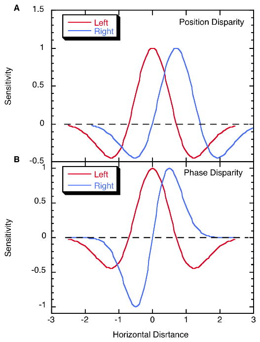
This essay reviews major developments - empirical and theoretical - in the field of binocular vision during the last 25years. We limit our survey primarily to work on human stereopsis, binocular rivalry and binocular contrast summation, with discussion where relevant of single-unit neurophysiology and human brain imaging. We identify several key controversies that have stimulated important work on these problems. In the case of stereopsis those controversies include position vs. phase encoding of disparity, dependence of disparity limits on spatial scale, role of occlusion in binocular depth and surface perception, and motion in 3D. In the case of binocular rivalry, controversies include eye vs. stimulus rivalry, role of "top-down" influences on rivalry dynamics, and the interaction of binocular rivalry and stereopsis. Concerning binocular contrast summation, the essay focuses on two representative models that highlight the evolving complexity in this field of study.


Figures

Figure 1
Figure 1
Horizontal receptive field profiles of vertically oriented left eye (red) and right eye (blue), receptive fields. A. A binocular unit combining responses of these two monocular units, which are identical except for a position shift, would be sensitive to a position disparity of approximately 0.8 horizontal distance units. B. Horizontal receptive field profiles of vertically oriented monocular receptive fields with even and odd symmetry. A binocular unit receiving excitation from these two would be sensitive to a 90° inter-ocular phase shift. Note that the two monocular profiles, in virtue of being phase shifted, have different optimal stimuli.
Figure 2
Figure 2
The geometry of occlusion and depth. A. Illustrates the case where one opaque rectangle positioned in front of another creates a monocular region M on the left for the left eye, and a similar monocular region M on the right for the right eye. B. This stereogram illustrates two partially occluded rectangles, one of which appears behind the large center rectangle, while the other appears in front and generates illusory contours. This clearly illustrates that depth is possible from occlusion. C. Depth ambiguity and constraint line under occlusion conditions. The solid front rectangle occludes the left eye's view of anything farther away than the dashed sight line. The right eye has an unoccluded view of the right edge of the rear solid rectangle along the solid sight line, but no depth information is available, so the rear black rectangle could lie anywhere along this sight line that is behind the left eye dashed occlusion line. Three possible locations are shown at A, B, and Panum. The latter is Panum s limiting case, the closest possible location of the occluded rectangle.
Figure 3
Figure 3
Panel a. Upper pair of figures are conventional rival stimuli, where separate, complete images are presented to the two eyes. Lower pair of figures are patch-wise rival stimuli that require interocular grouping in order for a coherent figure to be seen. (Figure reproduced with permission from Kovács et al, 1996. Copyright 1996, The National Academy of Sciences of the USA.). Panel b. Upper figure shows schematic of rapid eye-swap procedure used to induce stimulus rivalry. Rival targets are repetitively exchanged between the two eyes several times per second, with very rapid flicker used to mask transients associated with eye'swaps. Middle figure shows histograms of dominance durations measured without eye'swapping (left histogram) and with eye-swapping (right histogram). Lower figure summarizes the range of spatial and temporal frequencies yielding stimulus rivalry, measured using low and high contrast rival targets. (Figures in upper and lower panels reproduced with permission from Lee and Blake, 1999. Copyright 1999, Elsevier. Figure in middle panel reproduced with permission from Logothetis, Leopold and Sheinberg, 1996. Copyright Nature Press 1996.)
Figure 4
Figure 4
Panel a. Modulations in BOLD signal are correlated with reversals in rivalry state. Upper figure shows BOLD signals during dominance and suppression phases of binocular rivalry measured in human lateral geniculate nucleus in response to a high contrast pattern viewed by one eye and a low contrast pattern viewed by the other eye. Signals are time locked to transitions in perceptual state as indicated by observers perceptual tracking records. Graph on the left shows BOLD modulations before and immediately following transitions in rivalry state between the two rival patterns; graph on the right shows BOLD modulations before and immediately following physical removal and presentation of the two patterns that mimic alternations of rivalry. Middle figure shows the same as top figure, except that BOLD signals are being measured within voxels retinotopically localized within visual area V1. Lower figure shows BOLD signals from two ventral stream areas selectively responsive to pictures of faces (fusiform face area: FFA) and pictures of houses (parahippocampal place area: PPA). Graph on the left shows changes in BOLD signals time-locked to transitions in rivalry dominance from the house to the face; graph on the right shows changes in BOLD signal time-locked to transitions in rivalry dominance from the face to the house. (Top and middle figures in a reproduced with permission from Wunderlich et al., 2005. Copyright 2005, Nature Press. Bottom figure reproduced with permission from Tong et al. 1998. Copyright 1998, Cell Press.) Panel b. Modulations in visual-evoked potentials (VEP, upper figure) and action potentials from individual neurons in the temporal lobe (lower figure) recorded from human observers experiencing binocular rivalry. (Upper figure reproduced with permission from Brown and Norcia, 1997. Copyright Elsevier, 1997. Lower figure reproduced with permission from Kreiman et al., 2002. Copyright 2002, The National Academy of Sciences of the USA).
Figure 5
Figure 5
Multistage models of binocular contrast summation. a. Ding and Sperling model consisting of two pairs of contrast gain control mechanisms, with each pair linked in reciprocal inhibition. The earlier stage gain control is governed by the total weighted contrast energy (TCE) and the later stage gain control is selective for orientation and spatial frequency. b. Two-stage model published by Baker and Meese (2007). It comprises contrast gain control (divisive suppression) on separate orientation-selective monocular channels followed by phase-dependent binocular interactions of left- and right-eye channels (both in-phase and anti-phase components).

References

    1. Adelson EH, Bergen JR. Spatiotemporal energy models for the perception of motion. Journal of the Optical Society of America, A. 1985;2:284–299. - PubMed
    1. Alais D, Blake R. Binocular rivalry and perceptual ambiguity. Boston: MIT Press; 2005.
    1. Alais D, Cass J, O’Shea RP, Blake R. Visual sensitivity underlying changes in visual consciousness. Current Biology. 2010;20:1362–1367. - PMC - PubMed
    1. Alais D, Lorenceau J, Arrighi R, Cass JR. Contour interactions between pairs of Gabors engaged in binocular rivalry reveal a map of the association field. Vision Research. 2006;46:1473–1487. - PubMed
    1. Alais D, Melcher D. Strength and coherence of binocular rivalry depends on shared stimulus complexity. Vision Research. 2007;47:269–279. - PubMed

Publication types