. 2010 Dec 15;194(1):144-53.
doi: 10.1016/j.jneumeth.2010.10.010. Epub 2010 Oct 15.

Convection-enhanced delivery and systemic mannitol increase gene product distribution of AAV vectors 5, 8, and 9 and increase gene product in the adult mouse brain

Affiliations

Convection-enhanced delivery and systemic mannitol increase gene product distribution of AAV vectors 5, 8, and 9 and increase gene product in the adult mouse brain

Nikisha Carty et al. J Neurosci Methods. .

Abstract

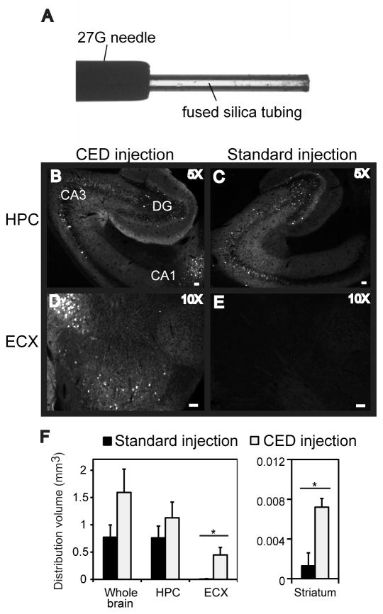
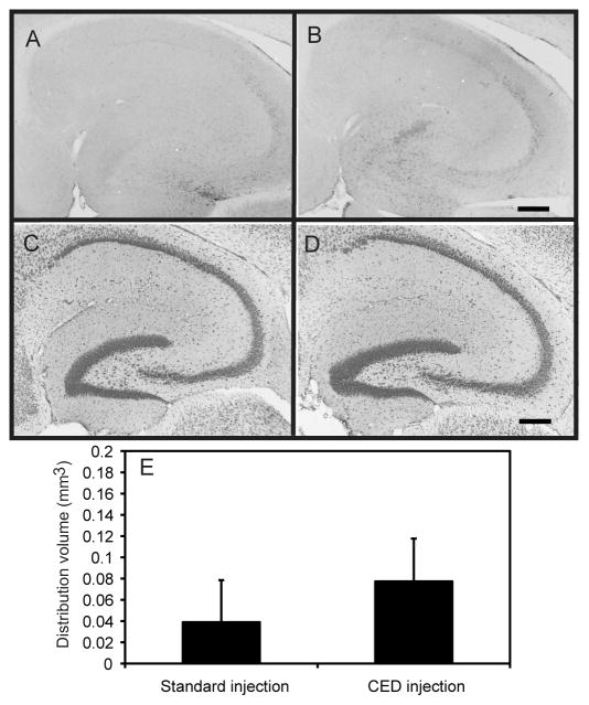
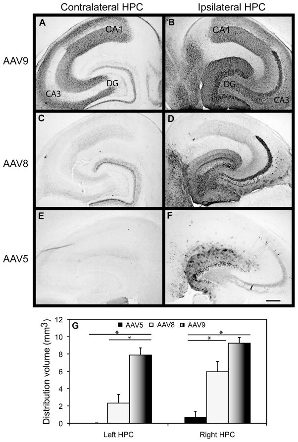
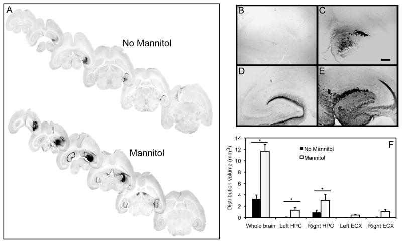
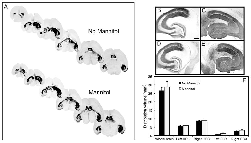
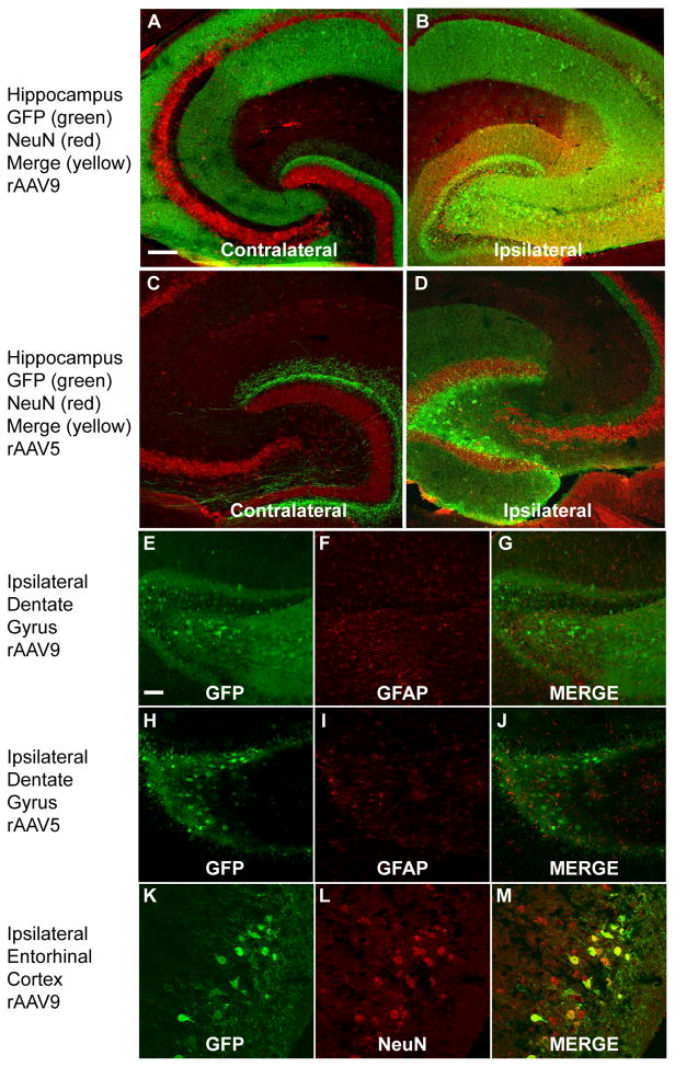
The use of recombinant adeno-associated viral (rAAV) vectors as a means of gene delivery to the central nervous system has emerged as a potentially viable method for the treatment of several types of degenerative brain diseases. However, a limitation of typical intracranial injections into the adult brain parenchyma is the relatively restricted distribution of the delivered gene to large brain regions such as the cortex, presumably due to confined dispersion of the injected particles. Optimizing the administration techniques to maximize gene distribution and gene expression is an important step in developing gene therapy studies. Here, we have found additive increases in distribution when 3 methods to increase brain distribution of rAAV were combined. The convection enhanced delivery (CED) method with the step-design cannula was used to deliver rAAV vector serotypes 5, 8 and 9 encoding GFP into the hippocampus of the mouse brain. While the CED method improved distribution of all 3 serotypes, the combination of rAAV9 and CED was particularly effective. Systemic mannitol administration, which reduces intracranial pressure, also further expanded distribution of GFP expression, in particular, increased expression on the contralateral hippocampi. These data suggest that combining advanced injection techniques with newer rAAV serotypes greatly improves viral vector distribution, which could have significant benefits for implementation of gene therapy strategies.


Figures

FIG. 1
FIG. 1
GFP expression is increased following intracranial administration of rAAV5 into the hippocampus using CED into 9-month-old mice. Panel A shows an image of the CED needle setup. The CED method (B, D, F) is compared to the standard injection method (C, E, F). Hippocampus (HPC), dentate gyrus (DG), cornu ammonis (CA) and entorhinal cortex (ECX) regions are shown. The volume of GFP fluorescence is graphed in panel G. The asterisk (*) indicates significance with a p-value < 0.05. The scale bar represents 120 μm for panels B, C, and 50 μm for panels D, E).
FIG. 2
FIG. 2
The CED method does not result in neuron loss or significant increase in CD45 expression. CD45 immunostaining in the hippocampus appears similar after the traditional injection method (panel A) or the CED injection method (panel B). No obvious loss of Nissl staining was observed after the traditional injection method (panel C) or the CED injection method (panel D). Quantification of CD45 immunostaining is represented as volume of stain in panel E. Scale bar = 120 μm for all panels.
FIG. 3
FIG. 3
Comparison of rAAV serotypes 5, 8, and 9 expressing GFP in the hippocampus (HPC). Images are presented depicting GFP expression in the contralateral HPC (A, C, E) and ipsilateral HPC (panels B, D, F) following a single CED intracranial injection into the right HPC with rAAV5, 8, or 9 serotypes. Quantification of the volume of positive staining for GFP is shown graphically in panel G. The asterisk (*) indicates significance with a p-value < 0.05. Scale = 120 μm. HPC= Hippocampus, DG= Dentate gyrus, CA=cornu ammonis.
FIG. 4
FIG. 4
GFP expression is increased following administration of rAAV5 using CED and systemic mannitol. Panel (A) shows stained sections from mannitol treated or untreated mice. Panels (B) and (C) show the left and right hippocampus respectively of a mouse injected with rAAV5 but no mannitol treatment. Panels (D) and (E) show the left and right hippocampus respectively of a mouse injected with rAAV5 and treated with mannitol. Panel (F) shows quantification of the volume of positive stain for anti-GFP immunohistochemistry. Hippocampus (HPC), entorhinal cortex (ECX) and whole brain (entire brain slice analyzed) are graphed. The asterisk (*) indicates significance with a p-value < 0.05. Scale = 120 μm.
FIG. 5
FIG. 5
GFP expression is increased following administration of rAAV9 using CED and systemic mannitol. Panel (A) shows stained sections from mannitol treated or untreated mice. Panels (B) and (C) show the left and right hippocampus respectively of a mouse injected with rAAV9 but no mannitol treatment. Panels (E) and (F) show the left and right hippocampus respectively of a mouse injected with rAAV9 and treated with mannitol. Panels (D) and (G) show the right entorhinal cortex of a mouse injected with rAAV9 not treated and treated with mannitol, respectively. Panel (H) shows quantification of the volume of positive stain for GFP. Hippocampus (HPC), entorhinal cortex (ECX) and whole brain (entire brain slice analyzed) are graphed. Scale = 120 μm (panels B–G).
FIG. 6
FIG. 6
Transduction efficiency and GFP expression in cell types following rAAV5 and rAAV9 administration into hippocampus. Panels (A) and (B) show the contralateral and ipsilateral hippocampus respectively of a mouse injected with rAAV9 and treated with mannitol. Red indicates Neu-N staining, green shows GFP fluorescence and yellow shows colocalization. Panels (C) and (D) show the contralateral and ipsilateral hippocampus respectively of a mouse injected with rAAV5 and treated with mannitol (staining as above panels). Panels (E–G) and (H–J) show no co-localization of astrocytic staining (GFAP) (panels F and I) and GFP fluorescence (panels E and H) in the dentate gyrus of the hippocampus for rAAV9 and rAAV5 respectively. Panels (K–M) show GFP fluorescence and Neu-N co-staining in the ipsilateral entorhinal cortex. The scale bar represents 120 μm in panels A–D and 50 μm in panels E–P.

References

    1. Akache B, Grimm D, Pandey K, Yant SR, Xu H, Kay MA. The 37/67-kilodalton laminin receptor is a receptor for adeno-associated virus serotypes 8, 2, 3, and 9. J Virol. 2006;80:9831–6. - PMC - PubMed
    1. Alisky JM, Hughes SM, Sauter SL, Jolly D, Dubensky TW, Jr, Staber PD, Chiorini JA, Davidson BL. Transduction of murine cerebellar neurons with recombinant FIV and AAV5 vectors. Neuroreport. 2000;11:2669–73. - PubMed
    1. Bankiewicz KS, Eberling JL, Kohutnicka M, Jagust W, Pivirotto P, Bringas J, Cunningham J, Budinger TF, Harvey-White J. Convection-enhanced delivery of AAV vector in parkinsonian monkeys; in vivo detection of gene expression and restoration of dopaminergic function using pro-drug approach. Exp Neurol. 2000;164:2–14. - PubMed
    1. Bantel Schaal U, zur Hausen H. Characterization of the DNA of a defective human parvovirus isolated from a genital site. Virology. 1984;134:52–63. - PubMed
    1. Bish LT, Morine K, Sleeper MM, Sanmiguel J, Wu D, Gao G, Wilson JM, Sweeney HL. Adeno-associated virus (AAV) serotype 9 provides global cardiac gene transfer superior to AAV1, AAV6, AAV7, and AAV8 in the mouse and rat. Hum Gene Ther. 2008;19:1359–68. - PMC - PubMed

Publication types

LinkOut - more resources