. 2010 Oct 19;18(4):329-40.
doi: 10.1016/j.ccr.2010.08.012.

Nuclear cyclin D1/CDK4 kinase regulates CUL4 expression and triggers neoplastic growth via activation of the PRMT5 methyltransferase

Affiliations

Nuclear cyclin D1/CDK4 kinase regulates CUL4 expression and triggers neoplastic growth via activation of the PRMT5 methyltransferase

Priya Aggarwal et al. Cancer Cell. .

Abstract

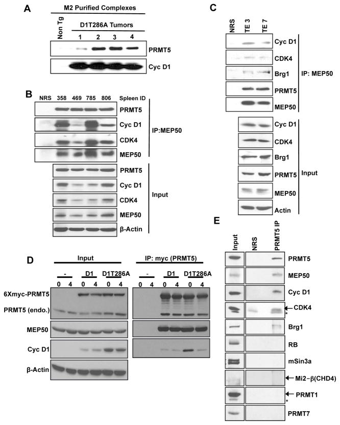
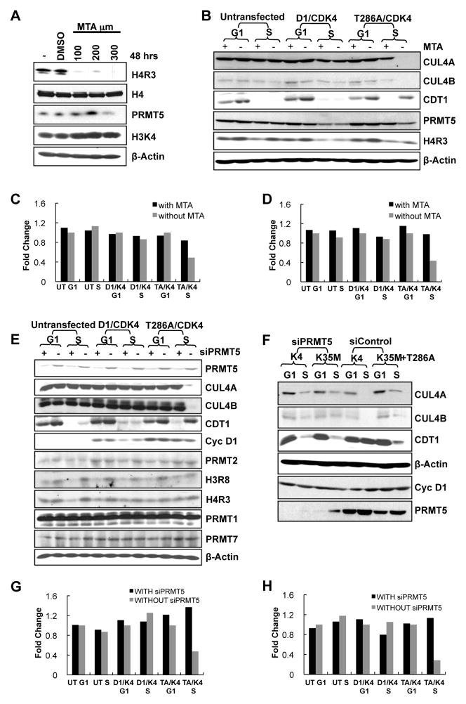
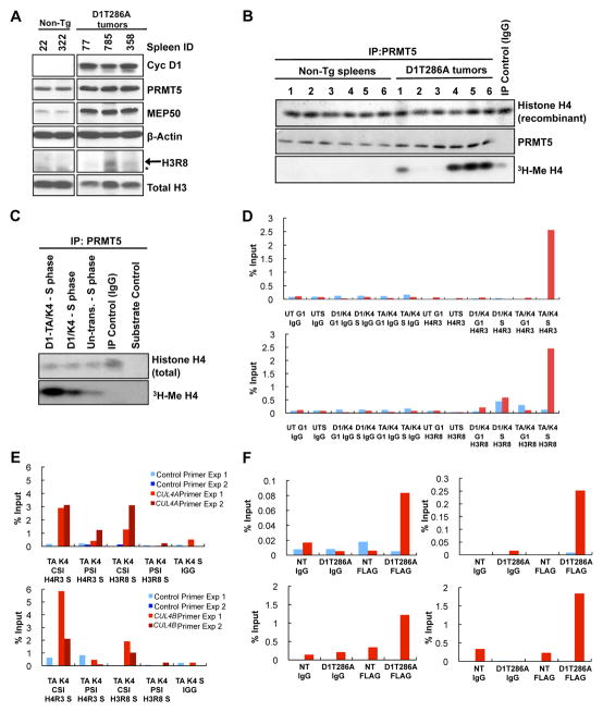
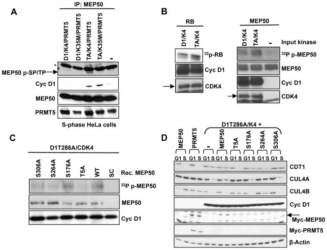
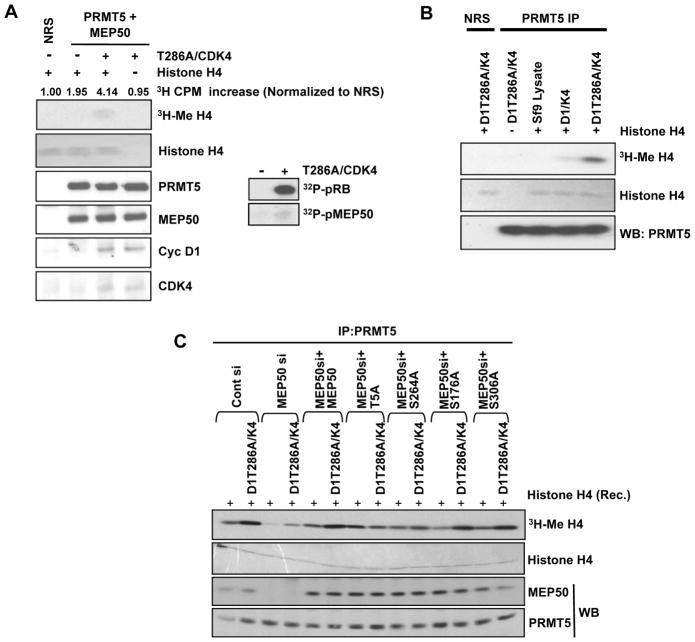
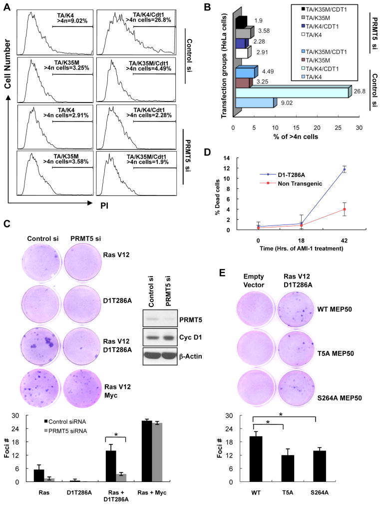
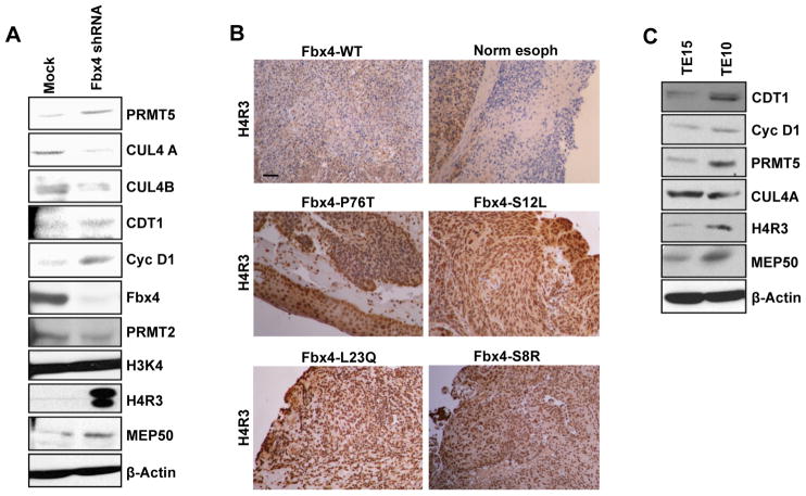
Cyclin D1 elicits transcriptional effects through inactivation of the retinoblastoma protein and direct association with transcriptional regulators. The current work reveals a molecular relationship between cyclin D1/CDK4 kinase and protein arginine methyltransferase 5 (PRMT5), an enzyme associated with histone methylation and transcriptional repression. Primary tumors of a mouse lymphoma model exhibit increased PRMT5 methyltransferase activity and histone arginine methylation. Analyses demonstrate that MEP50, a PRMT5 coregulatory factor, is a CDK4 substrate, and phosphorylation increases PRMT5/MEP50 activity. Increased PRMT5 activity mediates key events associated with cyclin D1-dependent neoplastic growth, including CUL4 repression, CDT1 overexpression, and DNA rereplication. Importantly, human cancers harboring mutations in Fbx4, the cyclin D1 E3 ligase, exhibit nuclear cyclin D1 accumulation and increased PRMT5 activity.


Figures

Figure 1
Figure 1. Identification of cyclin D1T286A/PRMT5/MEP50 complexes
(A) M2 (anti-Flag) purified complexes from Eμ-D1T286A (Flag epitope) transgenic tumors and non-transgenic spleens were blotted for PRMT5 and cyclin D1. (B) MEP50 immunoprecipitates (IP, 1mg whole cell lysate input) prepared from Eμ-D1T286A transgenic tumors were immunoblotted as indicated (C) MEP50 IP (1.5mg whole cell lysate input) from human esophageal cancer cell lines TE3, TE7 were immunoblotted as indicated (Top panel). Input lysates have been shown in the bottom panel. (D) HeLa cells transfected with either wild type cyclin D1 or D1T286A along with myc-PRMT5 and CDK4 were synchronized with aphidicolin. Cells were collected at 0 hours (G1/S boundary) and 4 hours (S-phase) after release. Myc-PRMT5 was pulled down using myc antibody (1mg whole cell lysate input) and immunoblotted as indicated (right panel), input lysates (left panel). (E) Cyclin D1/CDK4 complexes were isolated from Eμ-D1T286A lymphomas using M2-agarose followed by elution with Flag peptide. Eluted complexes were immunoprecipitated with normal rabbit serum (NRS) or PRMT5 antibody. 300μg of eluted complex served as input in NRS or PRMT5 IP, and western analysis was performed as indicated. See also Figure S1 and Table S1.
Figure 2
Figure 2. PRMT5/MEP50 mediates cyclin D1T286A/CDK4-dependent CUL4 repression and CDT1 stabilization
(A) HeLa cells were treated with MTA (5′ Deoxy-5″-methyl-thioadenosine) with concentrations ranging from 100 to 300 μM for 48 hours or vehicle DMSO. The cells were analyzed by western blot as indicated. (B) HeLa cells cultured for 24h in the absence or presence of 100μM MTA were transfected with vectors encoding cyclin D1 or D1T286A and CDK4. 24 hours post-transfection, cells were synchronized with nocodazole for 16–18 hrs; mitotic cells were then separated in two dishes. One was harvested 8h after release (G1-phase). Hydroxyurea was added to the second after release and harvested at 14h to obtain cells in S-phase; lysates were subjected to immunoblot as indicated. (C, D) RNA was collected from HeLa cells treated as in (B). The bars illustrate CUL4A (C) and CUL4B (D) mRNA levels with MTA treatment (first bar) and without MTA treatment (second bar) as analyzed by Real time PCR. One representative experiment of three biological independent experiments is presented. (E) HeLa cells were treated with or without siPRMT5 then transfected with wild type cyclin D1 or D1T286A plasmids along with CDK4. Cells were synchronized as in B, and immunoblotted as indicated. (F) HeLa cells were treated with siPRMT5 or si control smartpool for 24 hours, followed by transfection with cyclin D1T286A along with CDK4 or kinase dead CDK4(K35M) plasmids. Cells were synchronized as in (B), and lysates were immunoblotted as indicated. (G, H) Same as (C-D), except that cells were analyzed with and without siPRMT5 treatment. See also Figure S2.
Figure 3
Figure 3. Cyclin D1T286A/CDK4 activity increases PRMT5 methyltransferase activity in vivo
(A) Non-transgenic spleen or cyclin D1T286A-driven splenic lymphoma lysates were immunoblotted as indicated. (B) Methyltransferase activity of tumor-derived PRMT5 using recombinant histone H4 as the substrate. (C) Same as (B), except that PRMT5 complexes were immuno-purified from HeLa cells transfected with the indicated plasmids and synchronized in S-phase. (D-F) ChIP primer sets (1,2 and 3) were designed to span 1000–2000bp upstream of the first coding exon of either CUL4A or CUL4B respectively (primer design, Figure S3D). Primer set 3 was used for both CUL4A and CUL4B ChIP assays. Control primer sets were designed ~5000bp upstream of first coding exon of CUL4A/B. The CUL4A/B promoter primer (red bar) and control primer (blue bar) are displayed in all graphs. ChIP was performed for CUL4A (Figure 3D) and CUL4B (Figure S3E) using antibodies directed to di-methyl H4R3 (Figure 3D, top panel), H3R8 (Figure 3D, bottom panel; Figure S3E right panel), or normal mouse IgG (first 6 bars of each graph), on chromatin prepared from synchronized HeLa cells expressing wild type cyclin D1/CDK4, D1T286A/CDK4 or untransfected cells. DNA-protein immunoprecipitates and 5% of input chromatin were analyzed by qPCR for both CUL4A and CUL4B promoter regions. PCR values represent percentage of input. One representative experiment of three biological independent experiments is presented. (E) PRMT5 knockdown attenuates methylation of H4R3 and H3R8 of CUL4A (top graph) and CUL4B (bottom graph) promoter regions. ChIP was performed using di-methyl specific H4R3 and H3R8 antibodies as described in D; two representative experiments are shown. (F) Detection of cyclin D1T286A on the Cul4A/B proximal promoter by Flag ChIP from Eμ-D1T286A tumors. Two independent experiments are shown. See also Figure S3.
Figure 4
Figure 4. Cyclin D1/CDK4 kinase phosphorylates MEP50 in vitro and in vivo
(A) MEP50 was precipitated from HeLa cells transfected with vectors encoding cyclin D1, D1T286A along with either CDK4 or kinase dead CDK4(K35M) and synchronized in S-phase by sequential nocodazole and HU treatment. MEP50 phosphorylation was assessed using a commercially available phospho-SP/TP antibody. The arrow indicates the mobility of phospho-MEP50, which migrates faster than the (*) indicated non-specific immunoreactive band. Total MEP50, PRMT5 and cyclin D1 present in immune complexes were assessed using appropriate antibodies. (B) Phosphorylation of recombinant MEP50 or RB by cyclin D1/CDK4 or D1T286A/CDK4. Substrate phosphorylation was assessed by SDS-PAGE followed by autoradiography. (C) Cyclin Cyclin D1T286A/CDK4 phosphorylation of MEP50 at Thr-5, Ser-264 and Ser-306 in vitro. (D) Acute expression of MEP50 mutants Thr-5A and Ser-264A inhibit cyclin D1T286A/CDK4-dependent repression of CUL4 and CDT1 stabilization. See also Figure S4.
Figure 5
Figure 5. Cyclin D1T286A/CDK4 increases PRMT5 methyltransferase activity through MEP50
(A, B) Purified cyclin D1/CDK4 and D1T286A/CDK4 kinases increase catalytic activity of PRMT5/MEP50 in vitro. Purified cyclin/CDK4 complexes from Sf9 cells were mixed with purified recombinant PRMT5/MEP50 produced in Sf9 cells (A) or PRMT5 complexes purified from HeLa cells (B) and incubated for 30 min with ATP at 30°C. PRMT5 complexes were washed and methyltransferase activity assessed using 3H-Me (SAM) and recombinant Histone H4. (C) Same as (B), except PRMT5 was purified from HeLa cells following MEP50 knockdown and re-expression of the indicated MEP50 mutants.
Figure 6
Figure 6. PRMT5 knockdown inhibits cyclin D1T286A-dependent DNA re-replication, cell transformation and increases death of tumor cells
(A) Following transfection of HeLa cells with the indicated expression plasmids, TA (D1T286A), K4 (CDK4), K35M (CDK4(K35M)), CDT1, cell cycle profile was assessed by FACS after PI staining. (B) Graphical representation of the flow cytometry data from (A). The bars show the percentage of cells showing >4N DNA content. (C) Foci formation assay was performed by concurrent knockdown of murine PRMT5 and overexpression of the indicated proteins in NIH3T3 cells. Cells were grown for 14 days and stained with Giemsa to visualize foci. Quantification of data has been shown. Error bars represent ±SD and * indicates p-value < 0.05. (D) Single cell suspensions of splenocytes prepared from non-transgenic or malignant Eμ-D1T286A transgenic tumor burdened spleens were treated with 200μM arginine methyltransferase inhibitor AMI-1. The cells were analyzed for the inhibitor sensitivity by measuring the % cell death via AnnexinV –PI staining/flow cytometry. Values represent the mean of three independent experiments with error bars indicating ±SD. (E) Foci formation assay was performed by transfection of the indicated MEP50 constructs along with empty vector control or RasV12 plus cyclin D1T286A in NIH 3T3 cells. Cells were grown for 14 days and foci were analyzed as in (C). See also Figure S5.
Figure 7
Figure 7. Knockdown of Fbx4 stabilizes nuclear cyclin D1 resulting in increased PRMT5-dependent histone methylation
(A) Direct western analysis of the protein lysates prepared from control NIH3T3 or NIH3T3 harboring Fbx4 knockdown. (B) Representative immunohistochemistry images showing di-methyl H4R3 staining of human esophageal tumor sections. Scale bar = 100μm. (C) Direct western analysis of the protein lysates prepared from TE15 and TE10 cell lines.

Similar articles

Cited by

References

    1. Aggarwal P, Lessie MD, Lin DI, Pontano L, Gladden AB, Nuskey B, Goradia A, Wasik MA, Klein-Szanto AJ, Rustgi AK, et al. Nuclear accumulation of cyclin D1 during S phase inhibits Cul4-dependent Cdt1 proteolysis and triggers p53-dependent DNA rereplication. Genes Dev. 2007;21:2908–2922. - PMC - PubMed
    1. Alt JR, Cleveland JL, Hannink M, Diehl JA. Phosphorylation-dependent regulation of cyclin D1 nuclear export and cyclin D1-dependent cellular transformation. Genes Dev. 2000;14:3102–3114. - PMC - PubMed
    1. Bani-Hani K, Martin IG, Hardie LJ, Mapstone N, Briggs JA, Forman D, Wild CP. Prospective study of cyclin D1 overexpression in Barrett’s esophagus: association with increased risk of adenocarcinoma. J Natl Cancer Inst. 2000;92:1316–1321. - PubMed
    1. Barbash O, Zamfirova P, Lin DI, Chen X, Yang K, Nakagawa H, Lu F, Rustgi AK, Diehl JA. Mutations in Fbx4 inhibit dimerization of the SCF(Fbx4) ligase and contribute to cyclin D1 overexpression in human cancer. Cancer Cell. 2008;14:68–78. - PMC - PubMed
    1. Bartkova J, Lukas J, Muller H, Lutzhoft D, Strauss M, Bartek J. Cyclin D1 protein expression and function in human breast cancer. Int J Cancer. 1994a;57:353–361. - PubMed

Publication types

MeSH terms