. 2010 Dec 1;70(23):9827-36.
doi: 10.1158/0008-5472.CAN-10-1671. Epub 2010 Oct 15.

Inhibition of ALK, PI3K/MEK, and HSP90 in murine lung adenocarcinoma induced by EML4-ALK fusion oncogene

Affiliations

Inhibition of ALK, PI3K/MEK, and HSP90 in murine lung adenocarcinoma induced by EML4-ALK fusion oncogene

Zhao Chen et al. Cancer Res. .

Abstract

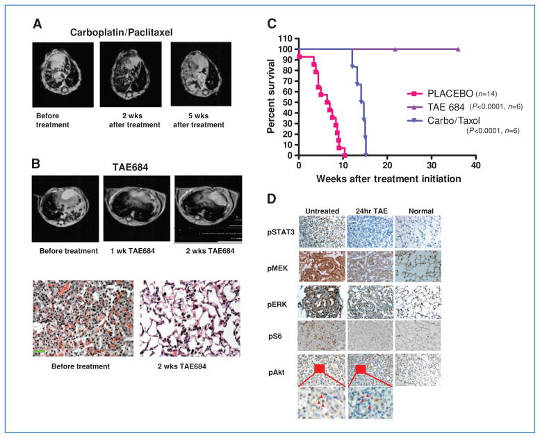
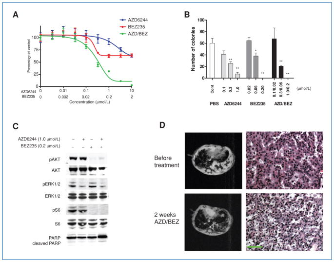
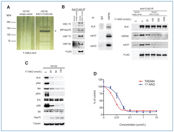
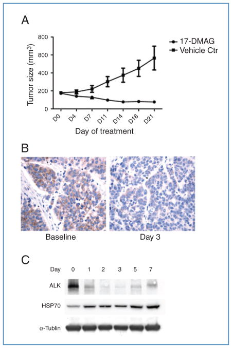
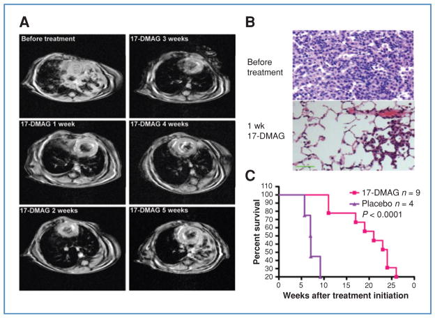
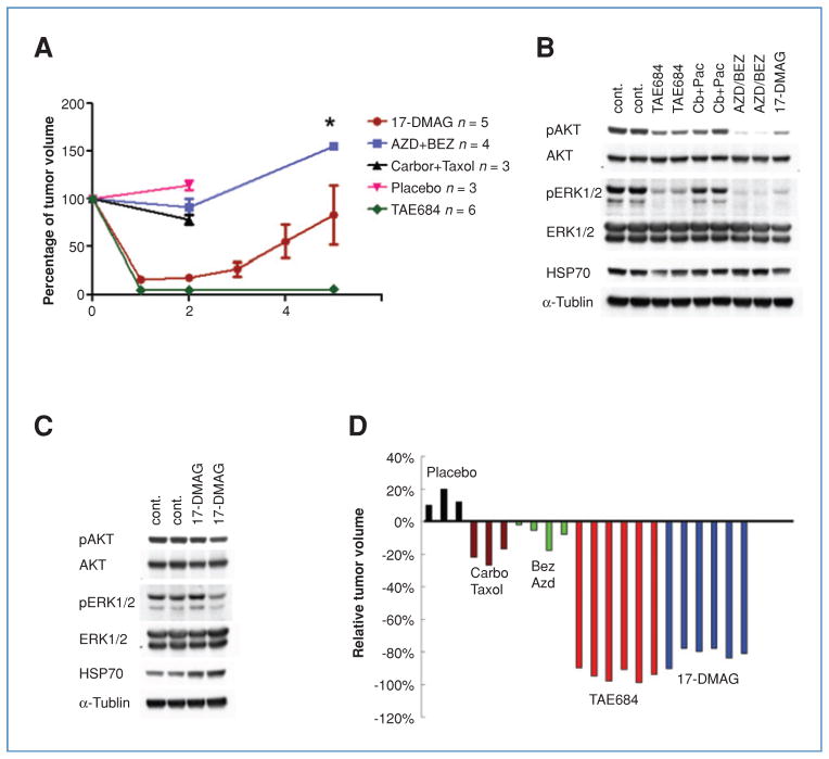
Genetic rearrangements of the anaplastic lymphoma kinase (ALK) kinase occur in 3% to 13% of non-small cell lung cancer patients and rarely coexist with KRASor EGFR mutations. To evaluate potential treatment strategies for lung cancers driven by an activated EML4-ALK chimeric oncogene, we generated a genetically engineered mouse model that phenocopies the human disease where this rearranged gene arises. In this model, the ALK kinase inhibitor TAE684 produced greater tumor regression and improved overall survival compared with carboplatin and paclitaxel, representing clinical standard of care. 18F-FDG-PET-CT scans revealed almost complete inhibition of tumor metabolic activity within 24 hours of TAE684 exposure. In contrast, combined inhibition of the PI3K/AKT and MEK/ERK1/2 pathways did not result in significant tumor regression. We identified EML4-ALK in complex with multiple cellular chaperones including HSP90. In support of a functional reliance, treatment with geldanamycin-based HSP90 inhibitors resulted in rapid degradation of EML4-ALK in vitro and substantial, albeit transient, tumor regression in vivo. Taken together, our findings define a murine model that offers a reliable platform for the preclinical comparison of combinatorial treatment approaches for lung cancer characterized by ALK rearrangement.


Conflict of interest statement

Disclosure of Potential Conflicts of Interest

No potential conflicts of interest were disclosed.

Figures

Figure 1
Figure 1
ALK-targeted therapy shows significant advantage over carboplatin/paclitaxel against murine EML-ALK lung cancer. A, representative MRI scans from a mouse harboring EML4-ALK–driven lung adenocarcinoma taken before treatment, and after 2 and 5 weeks of carboplatin/paclitaxel. B, MRI imaging and pathologic analysis demonstrate complete tumor regression following TAE684 for 2 weeks. Scale bar, 100 μm. C, survival curves following continuous treatment with vehicle, TAE684, or carboplatin/paclitaxel. D, tumor-bearing mice untreated or treated with TAE684 for 24 hours. Tumors were harvested and analyzed by immunohistochemistry for the indicated phosphoproteins, demonstrating significant downregulation of ALK signaling pathways. Small areas (insets) of pAkt staining are enlarged to compare signals before and after TAE684 treatment. Red arrows, tumor cells. Staining and histology of lungs from nontumor bearing mice are shown for comparison.
Figure 2
Figure 2
PI3K/mTOR and MEK/ERK1/2 inhibition reduce viability of EML4-ALK-dependent lung cancer cells in vitro but not in vivo. A, H3122 cells were treated with the indicated concentrations of the MEK inhibitor AZD6244, the PI3K-mTOR inhibitor BEZ235, or the combination of both drugs. Cell viability was determined after 72 hours by MTS assay. Data are presented as the percentage of viable cells compared with untreated cells. B, H3122 cells were treated with indicated doses of AZD6244, BEZ235, or both for 14 days, and colonies counted. Bars denote SD. Student’s t tests were performed comparing PBS with AZD6244, BEZ235, or AZD/BEZ. *, P < 0.01, **, P < 0.001. C, H3122 cells were treated with PBS, AZD6244 (1 μmol/L), BEZ235 (0.2 μmol/L), or both for 6 hours. Lysates were subjected to Western blotting with the indicated antibodies, demonstrating expected effects on signal transduction by these compounds. D, EML4-ALK tumor-bearing mice were treated with the combination of AZD6244 and BEZ235. Tumor volumes were documented by MRI imaging. Mice were sacrificed after 2 weeks of treatment for pathologic analysis. Scale bar, 100 μm.
Figure 3
Figure 3
Association of EML4-ALK proteins with the HSP complex. A, silver staining of Tandem Affinity-purified proteins associated with EML4-ALK V1. *, EML4-ALK; other bands represent novel proteins. B, EML4-ALK is a sensitive HSP90 client. Left, validation of EML4-ALK–associated proteins. Lysates from H3122 cells expressing FLAG-HA–tagged EML4-ALK V1 or empty vector were subjected to immunoprecipitation with anti-FLAG antibodies followed by Western blotting with antibodies against the indicated proteins, demonstrating the association of multiple HSP family members with ectopic EML4-ALK. Middle, association between endogenous EML4-ALK and HSP90 in H3122 cells. Lysates were subjected to immunoprecipitation with IgG or an anti-HSP90 antibody followed by Western blotting for ALK, cdc37, and p23. Right, the association of EML4-ALK with HSP90 and cdc37 is disrupted by 17-AAG treatment. Cells were treated with the indicated concentrations of 17-AAG for 1 hour. Lysates were subjected to immunoprecipitation with an anti-FLAG antibody followed by Western blotting for the indicated proteins. C, H3122 cells were treated with the indicated concentrations of 17-AAG for 24 hours and lysates analyzed by Western blotting for the indicated proteins, demonstrating depletion of ALK and downstream signaling proteins at a 17-AAG concentration as low as 25 nmol/L. D, H3122 cells were treated with TAE684 or 17-AAG for 72 hours, and analyzed using the MTS assay.
Figure 4
Figure 4
17-DMAG–mediated HSP90 inhibition is effective against EML4-ALK–driven lung cancer in H3122 xenograft. A, mice bearing H3122 xenografts were treated with vehicle or 17-DMAG, and tumor volumes were plotted over time. Bars denote SE. B, immunohistochemical analysis for ALK. Left, H3122 xenografts express EML4-ALK in the cytoplasm. Right, following 3 days of treatment with 17-DMAG, the expression of ALK was markedly reduced. C, Western blots of tumor lysates from mice treated with 17-DMAG at the indicated time points, demonstrating reduced ALK expression and induction of HSP70. Lane 0, untreated.
Figure 5
Figure 5
17-DMAG is effective against EML4-ALK–driven lung cancer in genetically engineered mouse model. A, representative MRI images from mouse 292, harboring EML4-ALK lung adenocarcinoma, treated for 5 consecutive weeks with 17-DMAG, demonstrating an initial response that was not durable (see Supplementary Table S4 for quantification of tumor volumes). B, hematoxylin and eosin (H&E) staining of tumor sections from mice left untreated or treated with 17-DMAG for 1 week, demonstrating only small residual tumor and restoration of normal lung architecture. Scale bar, 100 μm. C, Kaplan–Meier survival analysis of mice with EML4-ALK–driven lung cancer treated with 17-DMAG or placebo. Median survival of placebo control is 7.05 weeks whereas that for the 17-DMAG treated group is 21 weeks after treatment initiation.
Figure 6
Figure 6
Comparison of different therapeutic strategies in the murine EML4-ALK lung cancer model. A, dynamics of tumor volume change with different treatment strategies. Time points for tumor volume sampling are indicated by colored dots. Bars denote SE. *, n = 2, no SE calculated. B, comparison of ALK downstream signaling in mice with EML4-ALK–driven lung cancer treated for 1 day with the indicated drug(s). Tumor lysates were analyzed by Western blot with the indicated antibodies. C, ALK downstream signaling following 40 days of treatment with vehicle or 17-DMAG. Tumor lysates were analyzed by Western blot with the indicated antibodies. D, tumor volume changes by MRI (Image J software) 2 weeks after the indicated treatments. Each color represents a different treatment approach; each bar represents tumor volume change after treatment in 1 mouse.

References

    1. Morris SW, Kirstein MN, Valentine MB, Dittmer KG, Shapiro DN, Saltman DL, et al. Fusion of a kinase gene, ALK, to a nucleolar protein gene, NPM, in non-Hodgkin’s lymphoma. Science. 1994;263:1281–4. - PubMed
    1. Palmer RH, Vernersson E, Grabbe C, Hallberg B. Anaplastic lymphoma kinase: signalling in development and disease. Biochem J. 2009;420:345–61. - PMC - PubMed
    1. Koivunen JP, Mermel C, Zejnullahu K, Murphy C, Lifshits E, Holmes AJ, et al. EML4-ALK fusion gene and efficacy of an ALK kinase inhibitor in lung cancer. Clin Cancer Res. 2008;14:4275–83. - PMC - PubMed
    1. Perner S, Wagner PL, Demichelis F, Mehra R, Lafargue CJ, Moss BJ, et al. EML4-ALK fusion lung cancer: a rare acquired event. Neoplasia. 2008;10:298–302. - PMC - PubMed
    1. Shinmura K, Kageyama S, Tao H, Bunai T, Suzuki M, Kamo T, et al. EML4-ALK fusion transcripts, but no NPM-, TPM3-, CLTC-, ATIC-, or TFG-ALK fusion transcripts, in non-small cell lung carcinomas. Lung Cancer. 2008;61:163–9. - PubMed

Publication types

MeSH terms